. 2018 Aug 15:2018:7058953.
doi: 10.1155/2018/7058953. eCollection 2018.

Functional Connectivity Alterations in Children with Spastic and Dyskinetic Cerebral Palsy

Affiliations

Functional Connectivity Alterations in Children with Spastic and Dyskinetic Cerebral Palsy

Yun Qin et al. Neural Plast. .

Abstract

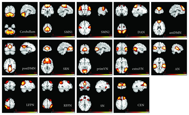
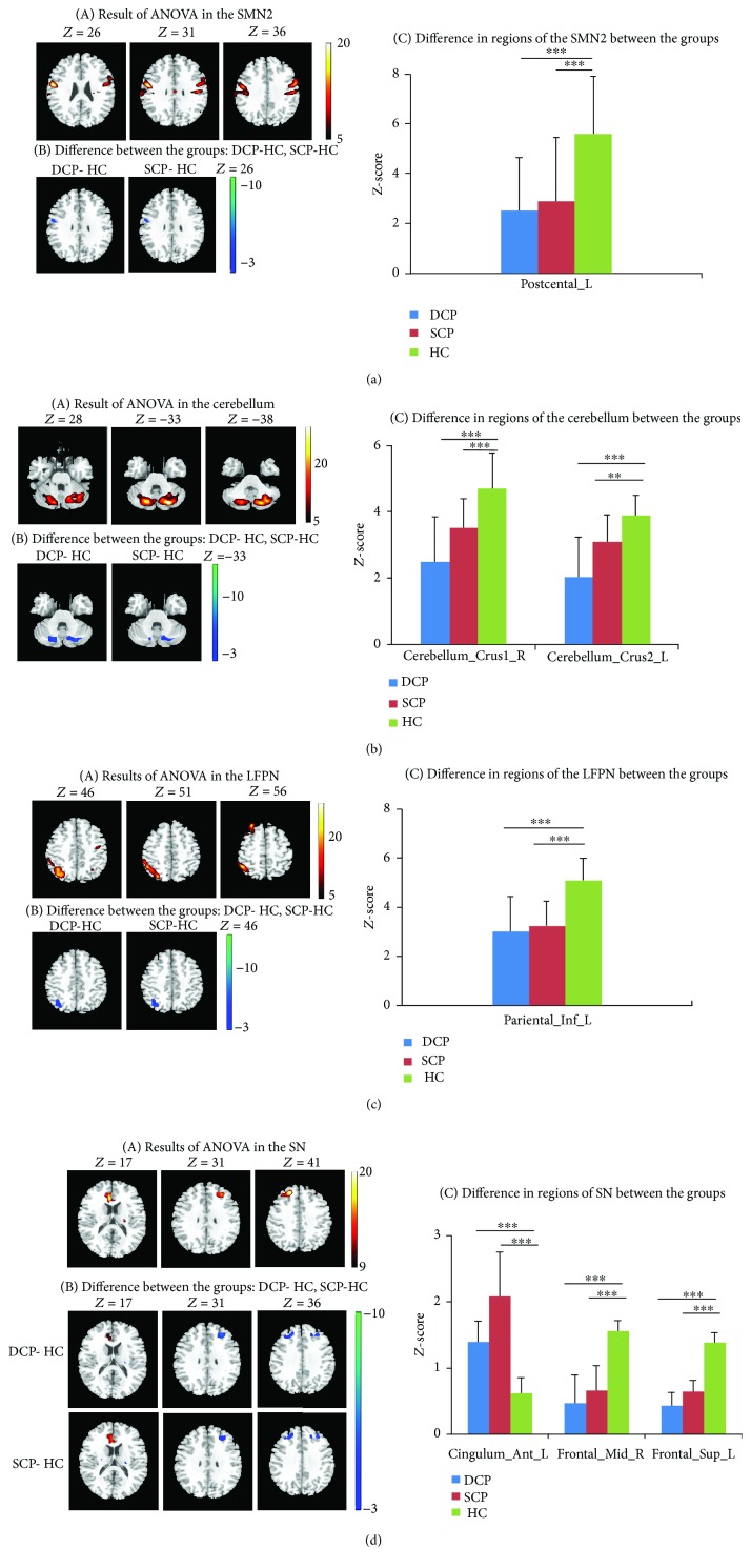
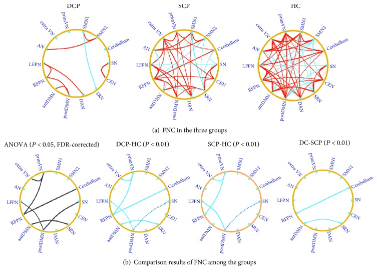
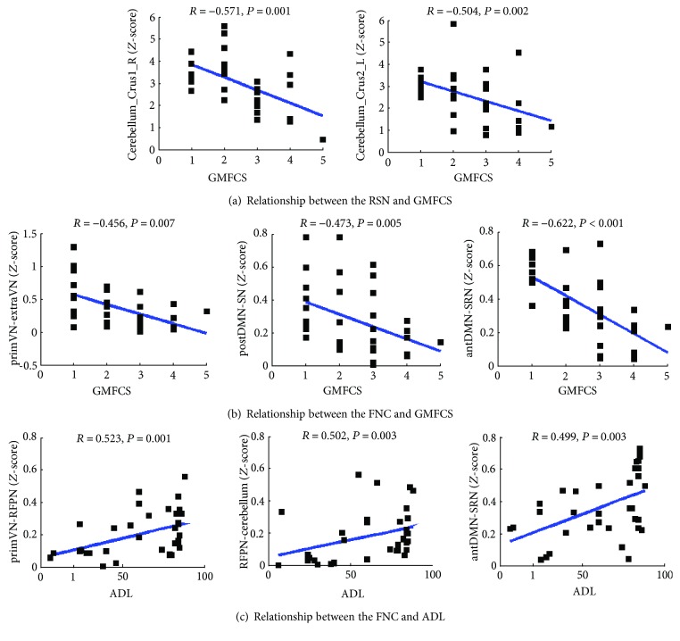
Cerebral palsy (CP) has long been investigated to be associated with a range of motor and cognitive dysfunction. As the two most common CP subtypes, spastic cerebral palsy (SCP) and dyskinetic cerebral palsy (DCP) may share common and distinct elements in their pathophysiology. However, the common and distinct dysfunctional characteristics between SCP and DCP on the brain network level are less known. This study aims to detect the alteration of brain functional connectivity in children with SCP and DCP based on resting-state functional MRI (fMRI). Resting-state networks (RSNs) were established based on the independent component analysis (ICA), and the functional network connectivity (FNC) was performed on the fMRI data from 16 DCP, 18 bilateral SCP, and 18 healthy children. Compared with healthy controls, altered functional connectivity within the cerebellum network, sensorimotor network (SMN), left frontoparietal network (LFPN), and salience network (SN) were found in DCP and SCP groups. Furthermore, the disconnections of the FNC consistently focused on the visual pathway; covariance of the default mode network (DMN) with other networks was observed both in DCP and SCP groups, while the DCP group had a distinct connectivity abnormality in motor pathway and self-referential processing-related connections. Correlations between the functional disconnection and the motor-related clinical measurement in children with CP were also found. These findings indicate functional connectivity impairment and altered integration widely exist in children with CP, suggesting that the abnormal functional connectivity is a pathophysiological mechanism of motor and cognitive dysfunction of CP.


Figures

Figure 1
Figure 1
The spatial patterns of 14 RSNs identified according to the group ICA in all children. SMN: sensorimotor network; DAN: dorsal attention network; antDMN: the anterior part of default mode network; postDMN: the posterior part of default mode network; SRN: the self-referential network; primVN: the primary visual network; extraVN: the extrastriate visual network; AN: the auditory network; LFPN: the left lateral frontoparietal network; RFPN: the right lateral frontoparietal network; SN: the salience network; CEN: the central executive network.
Figure 2
Figure 2
(a–d) Group comparison within four RSNs (SMN2, cerebellum, LFPN, and SN). (a) Significant difference was found in RSNs among the three groups. This result was achieved by performing one-way ANOVA on the maps of the three groups, with a threshold of corrected P < 0.05. (b) Differences were obtained between the DCP and HC group, as well as between the SCP and HC group, by performing post hoc test on the RSN maps (P < 0.001). (c) The bar maps present the between-group differences in the regions showing significant group difference. In the bar maps, ∗∗ P < 0.01, ∗∗∗ P < 0.001.
Figure 3
Figure 3
(a) The functional network connectivity in the DCP, SCP, and HC group. The red line represents the connection with positive correlation, and the blue line represents the connection with negative correlation. The results were obtained by the one-sample t-test with a threshold of corrected P < 0.05. (b) The comparison results of the functional network connectivity between the groups. The black line in the ANOVA result represents the altered connection among the three groups. Differences of functional network connectivity between DCP and HC and between SCP and HC, as well as between DCP and SCP, were observed. The blue solid line represents the connection with decreased positive FNC in CP children; and the blue dotted line represents the connection with decreased negative FNC in CP children.
Figure 4
Figure 4
(a) Relationship between cerebellum network and GMFCS scores. (b) Relationship between primVN-extraVN, postDMN-SN, antDMN-SRN connection, and GMFCS scores. (c) Relationship between primVN-RFPN, RFPN-cerebellum, antDMN-SRN connection, and ADL scores.

Similar articles

Cited by

References

    1. Novak I., Hines M., Goldsmith S., Barclay R. Clinical prognostic messages from a systematic review on cerebral palsy. Pediatrics. 2012;130(5):e1285–e1312. doi: 10.1542/peds.2012-0924. - DOI - PubMed
    1. Oskoui M., Coutinho F., Dykeman J., Jetté N., Pringsheim T. An update on the prevalence of cerebral palsy: a systematic review and meta-analysis. Developmental Medicine & Child Neurology. 2013;55(6):509–519. doi: 10.1111/dmcn.12080. - DOI - PubMed
    1. Bax M., Goldstein M., Rosenbaum P., et al. Proposed definition and classification of cerebral palsy, April 2005. Developmental Medicine and Child Neurology. 2005;47(8):571–576. doi: 10.1017/S001216220500112X. - DOI - PubMed
    1. Rosenbaum P. The definition and classification of cerebral palsy: are we any further ahead in 2006? NeoReviews. 2006;7(11):e569–e574. doi: 10.1542/neo.7-11-e569. - DOI
    1. Tomlin P. I. The StaticEncephalopathies. London: Times-Wolfe International; 1995.

Publication types