. 2018 Sep;16(3):2152-2159.
doi: 10.3892/etm.2018.6387. Epub 2018 Jul 2.

Reversible splenial lesion syndrome associated with acute Mycoplasma pneumoniae-associated encephalitis: A report of four cases and literature review

Affiliations

Reversible splenial lesion syndrome associated with acute Mycoplasma pneumoniae-associated encephalitis: A report of four cases and literature review

Xiaoyu Dong et al. Exp Ther Med. 2018 Sep.

Abstract

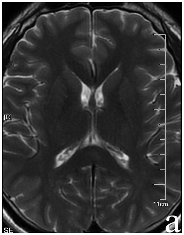
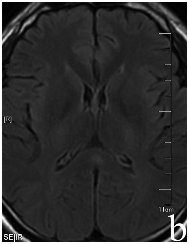
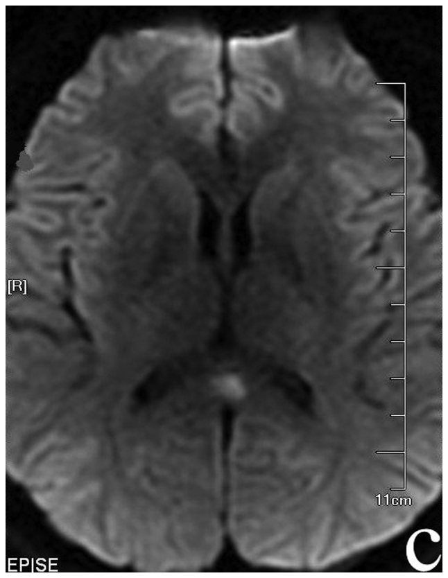
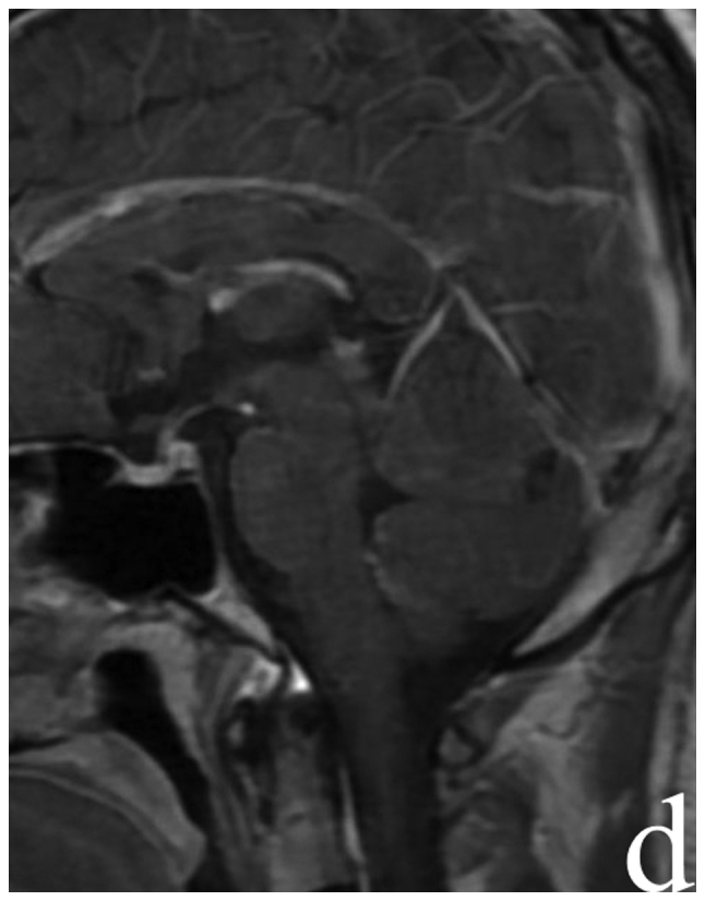
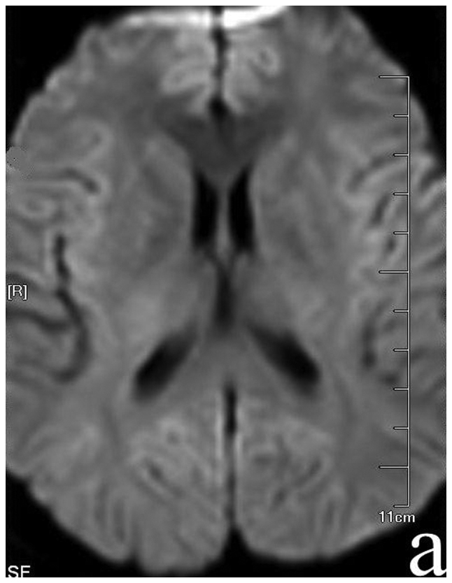
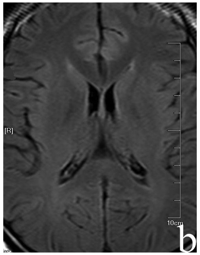
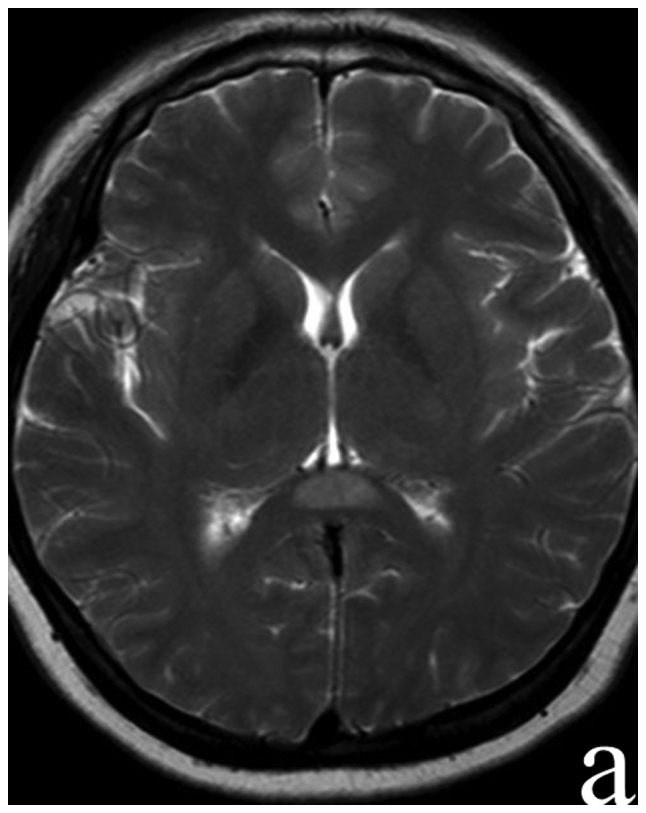
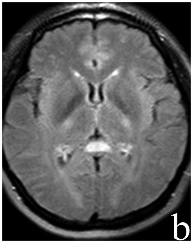
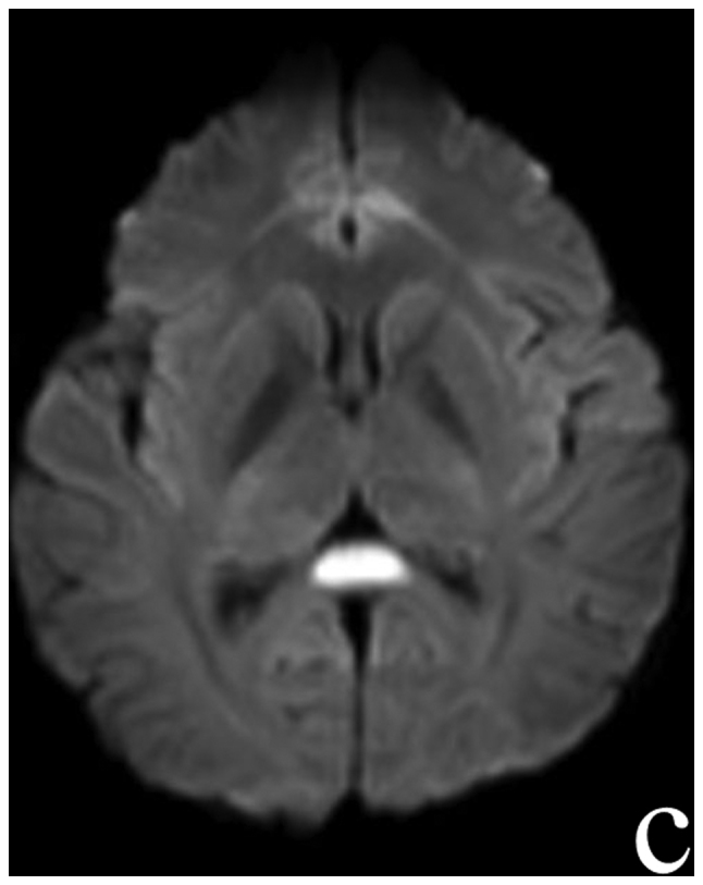
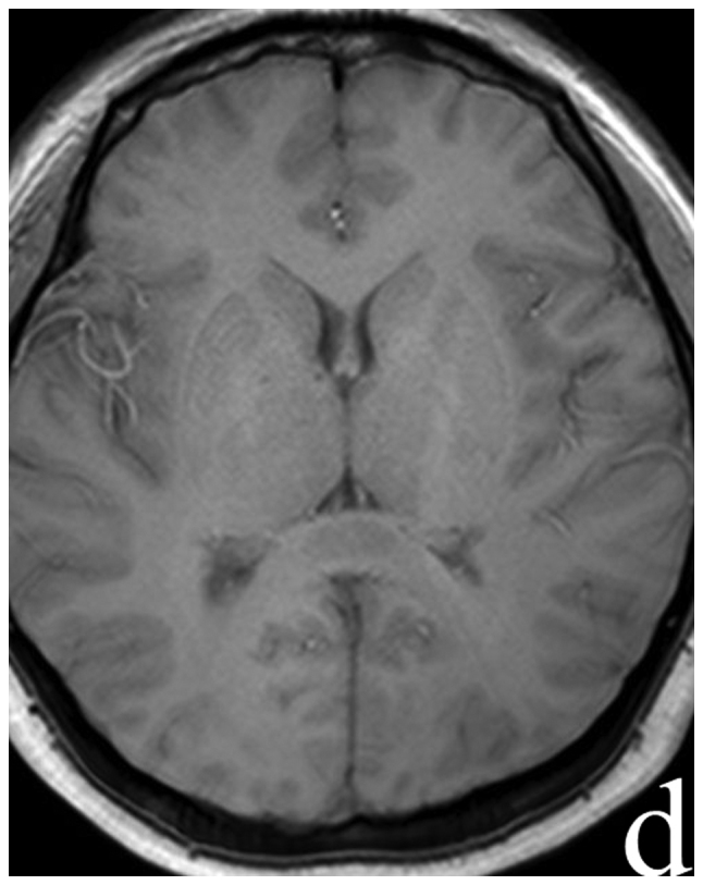
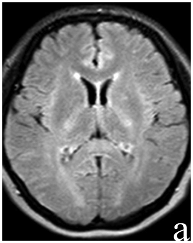
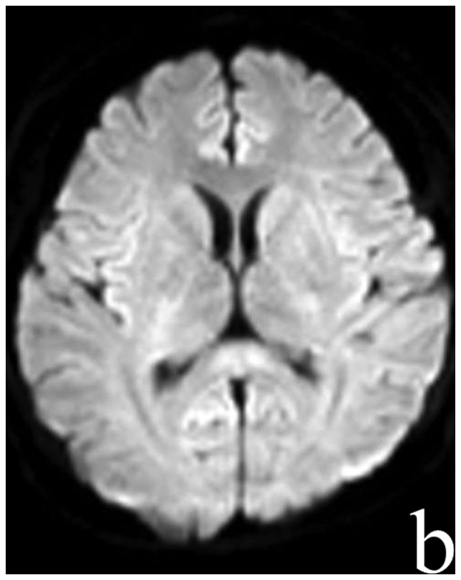
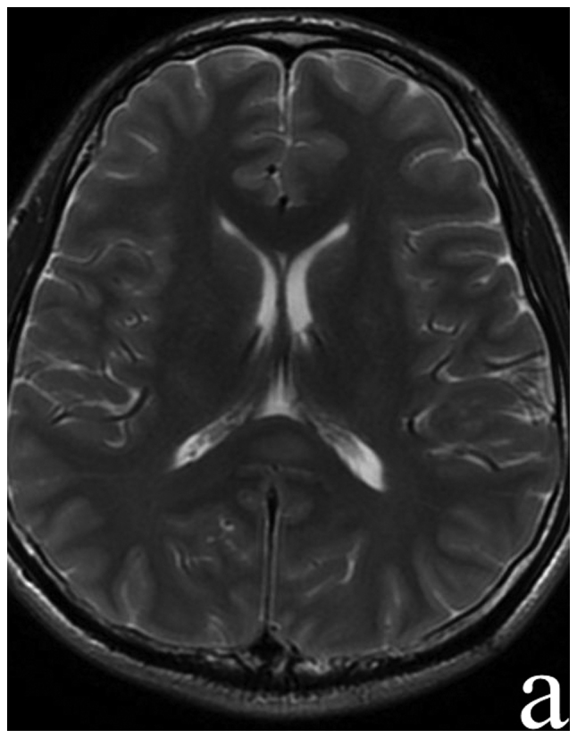
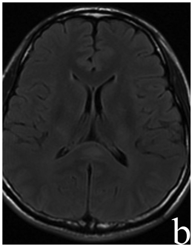
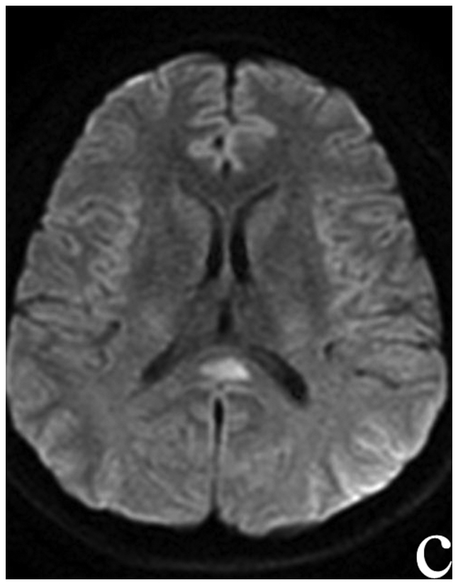
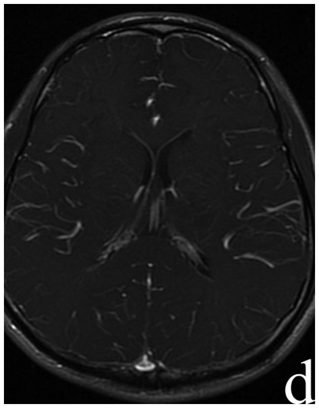
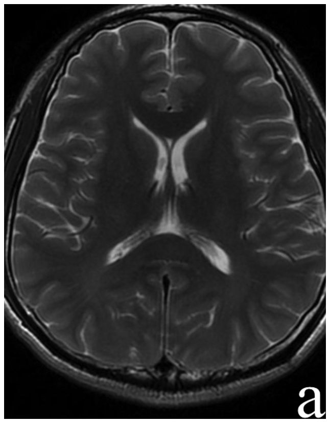
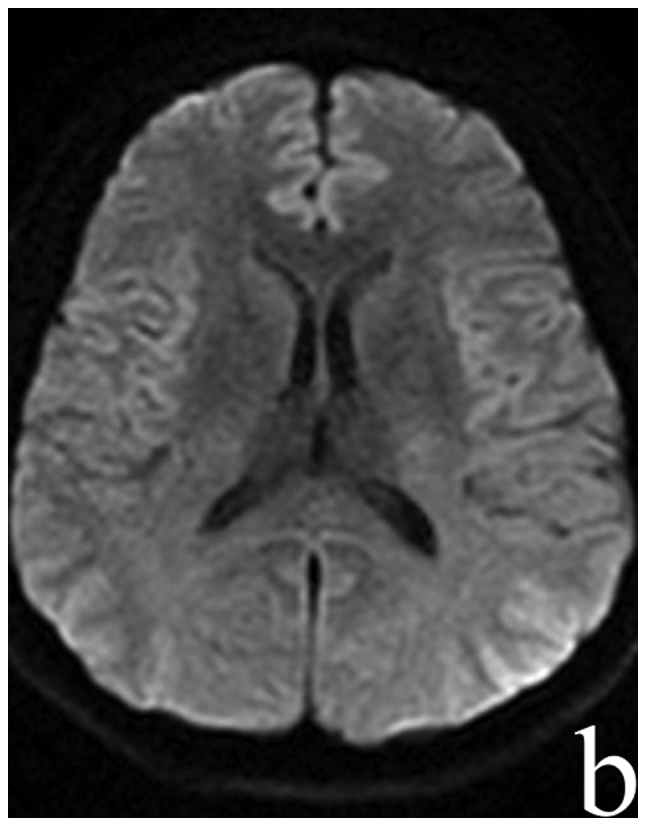
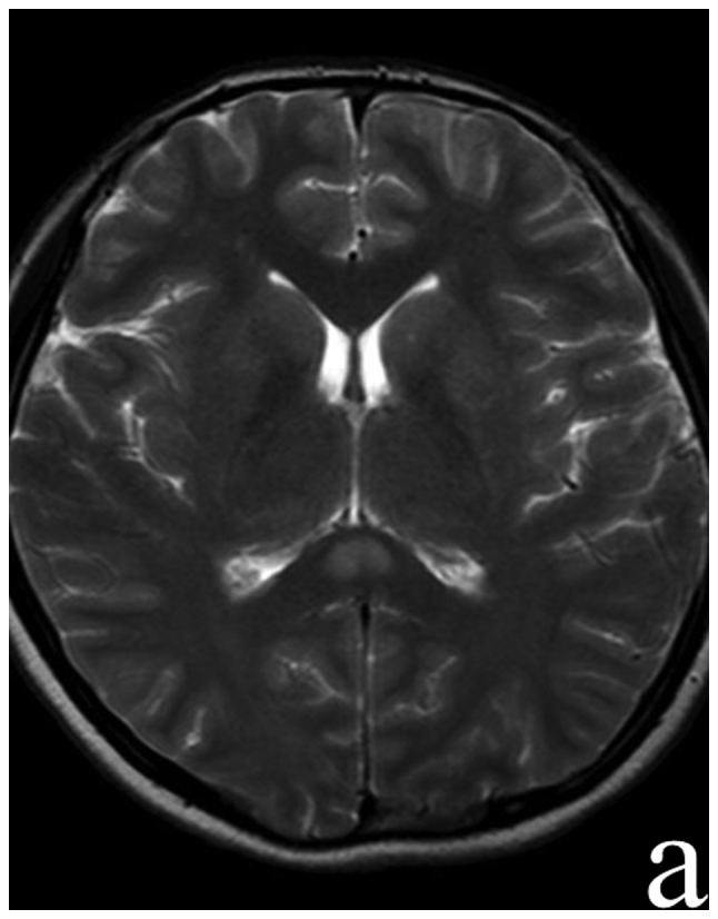
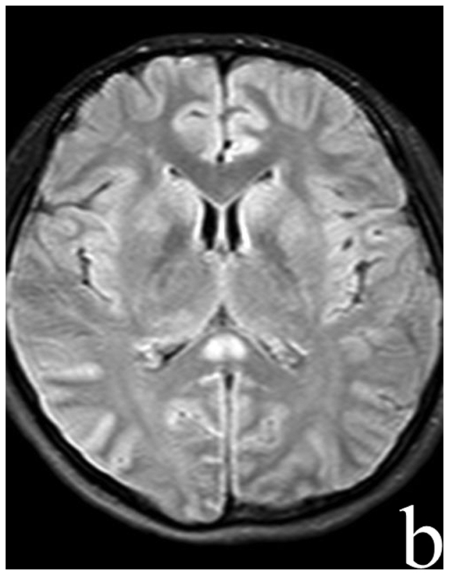
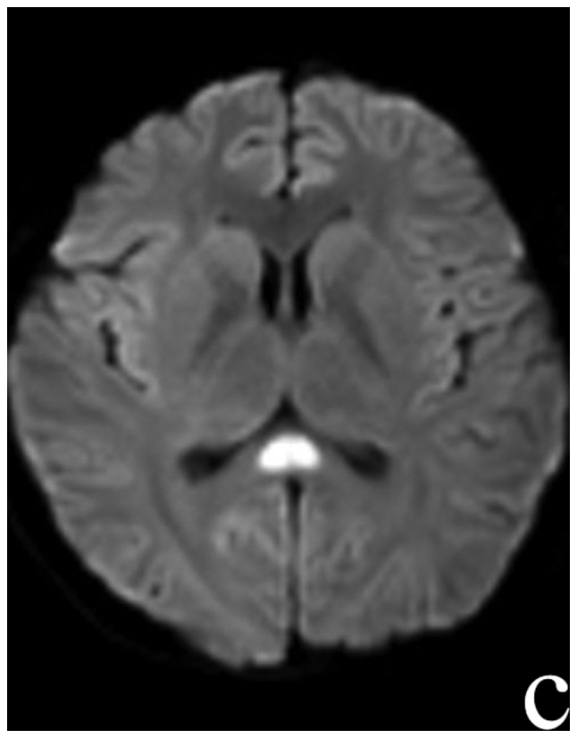
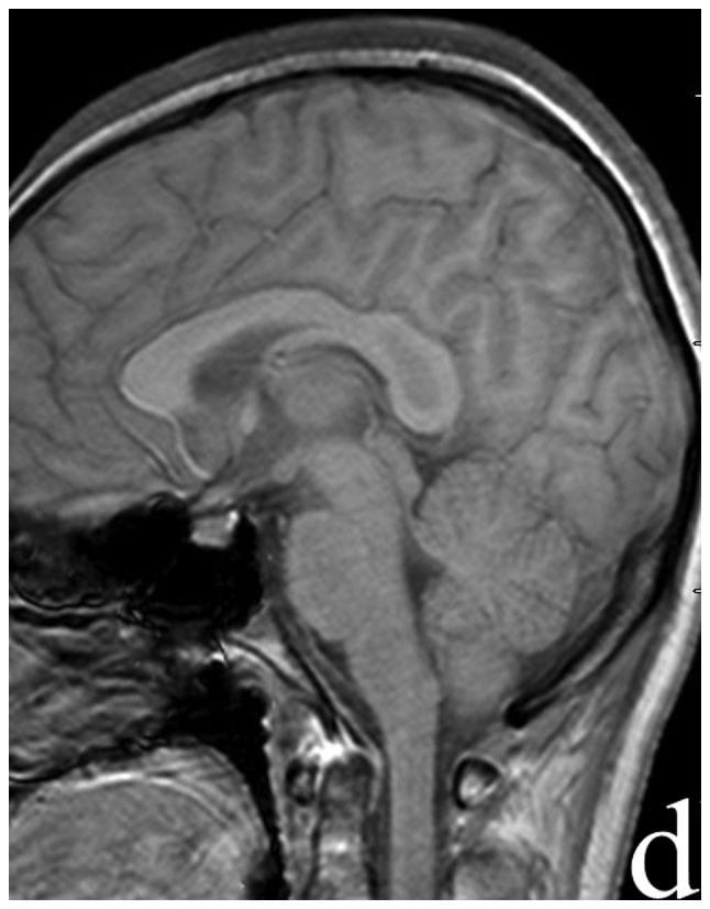
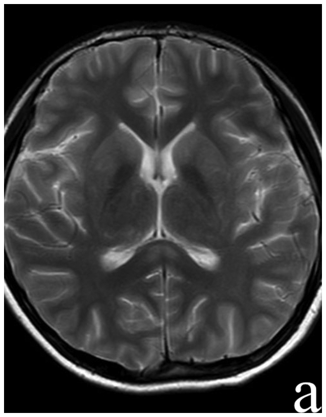
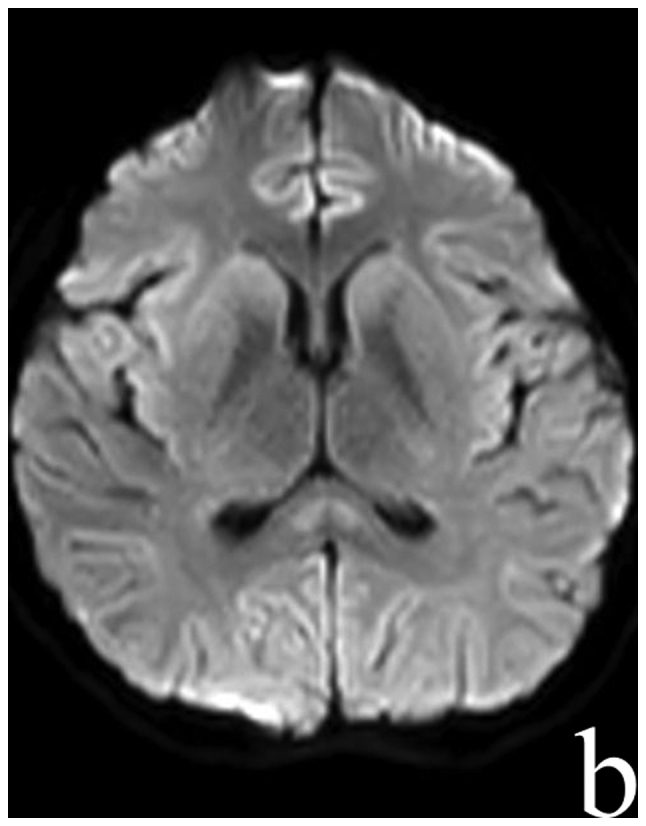
Reversible splenial lesion syndrome (RESLES) is a rare clinico-radiological syndrome that is defined as reversible lesions that involve the splenium of the corpus callosum (SCC). RESLES has been reported in patients with a broad spectrum of diseases and conditions, including infections, hypoglycemia and poisoning. The present report described four RESLES cases triggered by Mycoplasma pneumoniae (M. pneumoniae) and discussed the associated diagnostic challenges. Four cases of acute M. pneumoniae-associated encephalitis that displayed RESLES were reviewed. The clinical presentations were nonspecific in these patients. However, magnetic resonance imaging (MRI) revealed consistent lesions in the SCC with a hyperintensity in diffusion-weighted imaging (DWI) and hypointensities in T1WI, which disappeared after a variable lapse. Treatment with azithromycin or combined treatment with immunomodulatory agents if necessary led to a good prognosis. The present findings indicated that radiological diagnosis of RESLES should be considered in patients with M. pneumoniae-associated encephalitis. Furthermore, serum Mycoplasma antibody is important for the diagnosis of M. pneumoniae-associated encephalitis.

Keywords: Mycoplasma pneumoniae-associated encephalitis; magnetic resonance imaging; reversible splenial lesion syndrome; serum antibody.


Figures

Figure 1.
Figure 1.
Patient 1 MRI: (A) T2 W, (B) FLAIR, (C) DWI sequences showed hyperintensity signal in the splenium of corpus callosum, (D) sagittal T1 W enhanced scanning showed no enhancement effect in the lesion. The same region displayed hypointensity. MRI, magnetic resonance imaging; FLAIR, fluid attenuated inversion recovery; DWI, diffusion-weighted imaging.
Figure 1.
Figure 1.
Patient 1 MRI: (A) T2 W, (B) FLAIR, (C) DWI sequences showed hyperintensity signal in the splenium of corpus callosum, (D) sagittal T1 W enhanced scanning showed no enhancement effect in the lesion. The same region displayed hypointensity. MRI, magnetic resonance imaging; FLAIR, fluid attenuated inversion recovery; DWI, diffusion-weighted imaging.
Figure 2.
Figure 2.
Patient 1 follow-up MRI 10 days after admission. (A) DWI and (B) FLAIR sequences showed focal high-signal lesion in the splenial disappeared completely. MRI, magnetic resonance imaging; DWI, diffusion-weighted imaging; FLAIR, fluid attenuated inversion recovery.
Figure 2.
Figure 2.
Patient 1 follow-up MRI 10 days after admission. (A) DWI and (B) FLAIR sequences showed focal high-signal lesion in the splenial disappeared completely. MRI, magnetic resonance imaging; DWI, diffusion-weighted imaging; FLAIR, fluid attenuated inversion recovery.
Figure 3.
Figure 3.
Patient 2 MRI: (A) T2W, (B) FLAIR, (C) DWI sequences showed hyperintensity signal in the splenium of corpus callosum, and (D) T1 W showed the same region of hypointensity. MRI, magnetic resonance imaging; FLAIR, fluid attenuated inversion recovery; DWI, diffusion-weighted imaging.
Figure 3.
Figure 3.
Patient 2 MRI: (A) T2W, (B) FLAIR, (C) DWI sequences showed hyperintensity signal in the splenium of corpus callosum, and (D) T1 W showed the same region of hypointensity. MRI, magnetic resonance imaging; FLAIR, fluid attenuated inversion recovery; DWI, diffusion-weighted imaging.
Figure 4.
Figure 4.
Patient 2 follow up MRI 15 days after admission. (A) FLAIR and (B) DWI hyperintensities signal had disappeared completely in the splenial region. MRI, magnetic resonance imaging; FLAIR, fluid attenuated inversion recovery; DWI, diffusion-weighted imaging.
Figure 4.
Figure 4.
Patient 2 follow up MRI 15 days after admission. (A) FLAIR and (B) DWI hyperintensities signal had disappeared completely in the splenial region. MRI, magnetic resonance imaging; FLAIR, fluid attenuated inversion recovery; DWI, diffusion-weighted imaging.
Figure 5.
Figure 5.
Patient 3 MRI: (A) T2W, (B) FLAIR, (C) DWI sequences showed hyperintensity signal in the splenium of corpus callosum and (D) T1W enhanced scanning showed no enhancement effect. MRI, magnetic resonance imaging; FLAIR, fluid attenuated inversion recovery; DWI, diffusion-weighted imaging.
Figure 5.
Figure 5.
Patient 3 MRI: (A) T2W, (B) FLAIR, (C) DWI sequences showed hyperintensity signal in the splenium of corpus callosum and (D) T1W enhanced scanning showed no enhancement effect. MRI, magnetic resonance imaging; FLAIR, fluid attenuated inversion recovery; DWI, diffusion-weighted imaging.
Figure 6.
Figure 6.
Patient 3 follow up MRI 12 days after admission. (A) T2 and (B) DWI hyperintensities signal had disappeared completely in the splenial region. MRI, magnetic resonance imaging; DWI, diffusion-weighted imaging.
Figure 6.
Figure 6.
Patient 3 follow up MRI 12 days after admission. (A) T2 and (B) DWI hyperintensities signal had disappeared completely in the splenial region. MRI, magnetic resonance imaging; DWI, diffusion-weighted imaging.
Figure 7.
Figure 7.
Patient 4 MRI: (A) T2W, (B) FLAIR, (C) DWI sequences showed hyperintensity signal in the splenium of corpus callosum and (D) sagittal T1W sequence showed the same region of hypointensity. MRI, magnetic resonance imaging; FLAIR, fluid attenuated inversion recovery; DWI, diffusion-weighted imaging.
Figure 7.
Figure 7.
Patient 4 MRI: (A) T2W, (B) FLAIR, (C) DWI sequences showed hyperintensity signal in the splenium of corpus callosum and (D) sagittal T1W sequence showed the same region of hypointensity. MRI, magnetic resonance imaging; FLAIR, fluid attenuated inversion recovery; DWI, diffusion-weighted imaging.
Figure 8.
Figure 8.
Patient 4 follow up MRI 7 days after admission. (A) T2 and (B) DWI hyperintensities signal had decreased obviously in the splenial region. MRI, magnetic resonance imaging; DWI, diffusion-weighted imaging.
Figure 8.
Figure 8.
Patient 4 follow up MRI 7 days after admission. (A) T2 and (B) DWI hyperintensities signal had decreased obviously in the splenial region. MRI, magnetic resonance imaging; DWI, diffusion-weighted imaging.

References

    1. Lind K, Benzon MW, Jensen JS, Clyde WA., Jr A seroepidemiological study of Mycoplasma pneumoniae infections in Denmark over the 50-year period 1946–1995. Eur J Epidemiol. 1997;13:581–586. doi: 10.1023/A:1007353121693. - DOI - PubMed
    1. Bébéar C, Dupon M, Renaudin H, de Barbeyrac B. Potential improvements in therapeutic options for mycoplasmal respiratory infections. Clin Infect Dis. 1993;17(Suppl 1):S202–S207. doi: 10.1093/clinids/17.Supplement_1.S202. - DOI - PubMed
    1. Foy HM. Infections caused by Mycoplasma pneumoniae and possible carrier state in different populations of patients. Clin Infect Dis. 1993;17(Suppl 1):S37–S46. doi: 10.1093/clinids/17.Supplement_1.S37. - DOI - PubMed
    1. Narita M. Pathogenesis of neurologic manifestations of Mycoplasma pneumoniae infection. Pediatr Neurol. 2009;41:159–166. doi: 10.1016/j.pediatrneurol.2009.04.012. - DOI - PubMed
    1. Kuwahara M, Samukawa M, Ikeda T, Morikawa M, Ueno R, Hamada Y, Kusunoki S. Characterization of the neurological diseases associated with Mycoplasma pneumoniae infection and anti-glycolipid antibodies. J Neurol. 2017;264:467–475. doi: 10.1007/s00415-016-8371-1. - DOI - PubMed