Hypoxia-reoxygenation induced necroptosis in cultured rat renal tubular epithelial cell line
- PMID: 30186575
- PMCID: PMC6118090
- DOI: 10.22038/IJBMS.2018.26276.6444
Hypoxia-reoxygenation induced necroptosis in cultured rat renal tubular epithelial cell line
Abstract
Objectives: The aim of this study is to explore the potential role of hypoxia/reoxygenation in necroptosis in cultured rat renal tubular epithelial cell line NRK-52E, and further to investigate its possible mechanisms.
Materials and methods: Cells were cultured under different hypoxia-reoxygenation conditions in vitro. MTT assay was used to measure the cell proliferation of cells that were exposed to hypoxia-reoxygenation conditions at different time points. Receptor-interacting protein 1,3 (RIP1 and RIP3) and NF-κB were detected by Western-blot analysis. Co-immunoprecipitation (Co-IP) was conducted to investigate the formation of necrosome. Necrostatin-1 (Nec-1) was adopted to inhibit the occurrence of necroptosis. In addition, morphological changes of cells after hypoxia-reoxygenation interference were observed under transmission electron microscope (TEM).
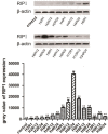
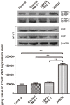
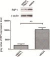
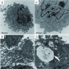
Results: MTT assay indicated that hypoxia-reoxygenation treatment can cause a decrease in cell viability. Particularly, 6 hr of hypoxia and 24 hr of reoxygenation (H6R24 group) resulted in the lowest cell viability. Western-blot results indicated that the expression of RIP3 significantly increased in H6R24 group while the expression of NF-κB is decreased. Co-IP results demonstrated that the interaction between RIP1 and RIP3 was stronger in the hypoxia-reoxygenation induced group than the other groups, furthermore, treatment with Nec-1 reduced the formation of necrosome. TEM observation results showed that hypoxia-reoxygenation treated cells showed typical morphological characteristics of necroptosis and autophagy.
Conclusion: Hypoxia-reoxygenation treatment can induce necroptosis in NRK-52E cells, and this effect can be inhibited by Nec-1. In addition, the mechanism of necroptosis induced by hypoxia-reoxygenation injury on cells may be related to the low expression of NF-κB.
Keywords: Cell line; Hypoxia-Reoxygenation Nec-1; Necrosome; Receptor interacting protein.
Figures


Similar articles
-
Detection of Necroptosis in Ligand-Mediated and Hypoxia-Induced Injury of Hepatocytes Using a Novel Optic Probe-Detecting Receptor-Interacting Protein (RIP)1/RIP3 Binding.Oncol Res. 2018 Apr 10;26(3):503-513. doi: 10.3727/096504017X15005102445191. Epub 2017 Jul 21. Oncol Res. 2018. PMID: 28770700 Free PMC article.
-
Resveratrol inhibits necroptosis by mediating the TNF-α/RIP1/RIP3/MLKL pathway in myocardial hypoxia/reoxygenation injury.Acta Biochim Biophys Sin (Shanghai). 2021 Mar 26;53(4):430-437. doi: 10.1093/abbs/gmab012. Acta Biochim Biophys Sin (Shanghai). 2021. PMID: 33686403
-
[Hypoxia/reoxygenation and lipopolysaccharide induced nuclear factor-ΚB and hypoxia-inducible factor-1α signaling pathways in intestinal epithelial cell injury and the interventional effect of emodin].Zhonghua Wei Zhong Bing Ji Jiu Yi Xue. 2014 Jun;26(6):409-14. doi: 10.3760/cma.j.issn.2095-4352.2014.06.009. Zhonghua Wei Zhong Bing Ji Jiu Yi Xue. 2014. PMID: 24912640 Chinese.
-
Heat stress induces RIP1/RIP3-dependent necroptosis through the MAPK, NF-κB, and c-Jun signaling pathways in pulmonary vascular endothelial cells.Biochem Biophys Res Commun. 2020 Jul 12;528(1):206-212. doi: 10.1016/j.bbrc.2020.04.150. Epub 2020 May 26. Biochem Biophys Res Commun. 2020. PMID: 32471717
-
Necrostatin-1 attenuates ischemia injury induced cell death in rat tubular cell line NRK-52E through decreased Drp1 expression.Int J Mol Sci. 2013 Dec 18;14(12):24742-54. doi: 10.3390/ijms141224742. Int J Mol Sci. 2013. PMID: 24351845 Free PMC article.
Cited by
-
Sirtuin 3 deficiency exacerbates diabetic cardiomyopathy via necroptosis enhancement and NLRP3 activation.Acta Pharmacol Sin. 2021 Feb;42(2):230-241. doi: 10.1038/s41401-020-0490-7. Epub 2020 Aug 7. Acta Pharmacol Sin. 2021. PMID: 32770173 Free PMC article.
-
Activation of Delta-Opioid Receptor Protects ARPE19 Cells against Oxygen-Glucose Deprivation/Reoxygenation-Induced Necroptosis and Apoptosis by Inhibiting the Release of TNF-α.J Ophthalmol. 2022 Nov 19;2022:2285663. doi: 10.1155/2022/2285663. eCollection 2022. J Ophthalmol. 2022. PMID: 36457949 Free PMC article.
-
Soluble receptor for advanced glycation end products protects from ischemia- and reperfusion-induced acute kidney injury.Biol Open. 2022 Jan 15;11(1):bio058852. doi: 10.1242/bio.058852. Epub 2022 Feb 4. Biol Open. 2022. PMID: 34812852 Free PMC article.
References
-
- Xiong K, Liao H, Long L, Ding Y, Huang J, Yan J. Necroptosis contributes to methamphetamine-induced cytotoxicity in rat cortical neurons. Toxicol In Vitro. 2016;3:163–168. - PubMed
-
- Himmelfarb J, Ikizler TA. Acute kidney injury: changing lexicography, definitions, and epidemiology. Kidney Int. 2007;71:971–976. - PubMed
-
- Jie Zhou CX, Wenwen Wang, Xinglu Fu, Liang Jinqiang, Yuwen Qiu, Jin Jin, Jingfen Xu, Zhiying Huang. Triptolide-induced oxidative stress involved with Nrf2 contribute to cardiomyocyte apoptosis through mitochondrial dependent pathways. Toxicol Lett. 2014;230:454–466. - PubMed
-
- Andreas Hartmann J-DT, Stéphane Hunot, Kristy Kikly, Baptiste A, Faucheux , Annick Mouatt-Prigent, Merle Ruberg, Yves Agid, Etienne C. Caspase-8 is an effector in apoptotic death of dopaminergic neurons in Parkinson’s disease, but pathway inhibition results in neuronal necrosis. J Neurosci . 2001;21:2247–2255. - PMC - PubMed
LinkOut - more resources
Full Text Sources
Research Materials
Miscellaneous
