

Gallstone ileus with spontaneous evacuation: A case report
- PMID: 30186731
- PMCID: PMC6119795
- DOI: 10.1002/jgf2.196
Gallstone ileus with spontaneous evacuation: A case report
Abstract
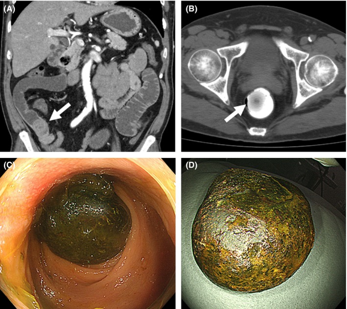
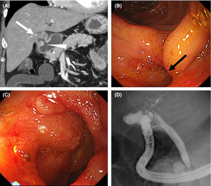
A 65-year-old man was referred to our hospital for abdominal pain. He had a history of enterotomy with stone extraction for gallstone ileus. On abdominal computed tomography, a stone measuring 32 × 28 mm lodged in the jejunum was identified. He was diagnosed with gallstone ileus and treated using a nasal ileus tube. Four days after admission, computed tomography showed that the stone had passed into the rectum. The gallstone was spontaneously evacuated on the same day. A fistula with the gallbladder was found in the duodenal bulb. The patient's condition improved, and he was discharged 9 days after admission.
Keywords: cholecystoduodenal fistula; gallstone; ileus; spontaneous evacuation.
Figures
References
-
- Clavien PA, Richon J, Burgan S, Rohner A. Gallstone ileus. Br J Surg. 1990;77:737–42. - PubMed
-
- Reisner RM, Cohen JR. Gallstone ileus: a review of 1001 reported cases. Am Surg. 1994;60:441–6. - PubMed
-
- Sesti J, Okoro C, Parikh M. Laparoscopic enterolithotomy for gallstone ileus. J Am Coll Surg. 2013;217:e13–5. - PubMed
-
- Chatterjee S, Chaudhuri T, Ghosh G, Ganguly A. Gallstone ileus–an atypical presentation and unusual location. Int J Surg. 2008;6:e55–6. - PubMed
Publication types
LinkOut - more resources
Full Text Sources
Other Literature Sources