. 2018 Aug 13;8(9):1524-1543.
doi: 10.1002/2211-5463.12497. eCollection 2018 Sep.

2D DIGE proteomic analysis reveals fasting-induced protein remodeling through organ-specific transcription factor(s) in mice

Affiliations

2D DIGE proteomic analysis reveals fasting-induced protein remodeling through organ-specific transcription factor(s) in mice

Shotaro Kamata et al. FEBS Open Bio. .

Abstract

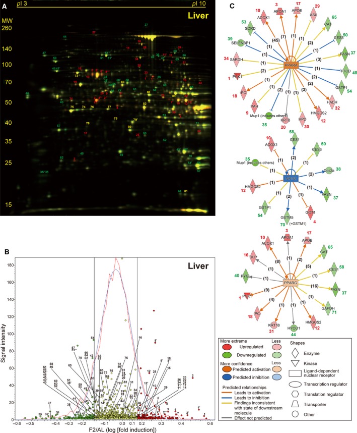
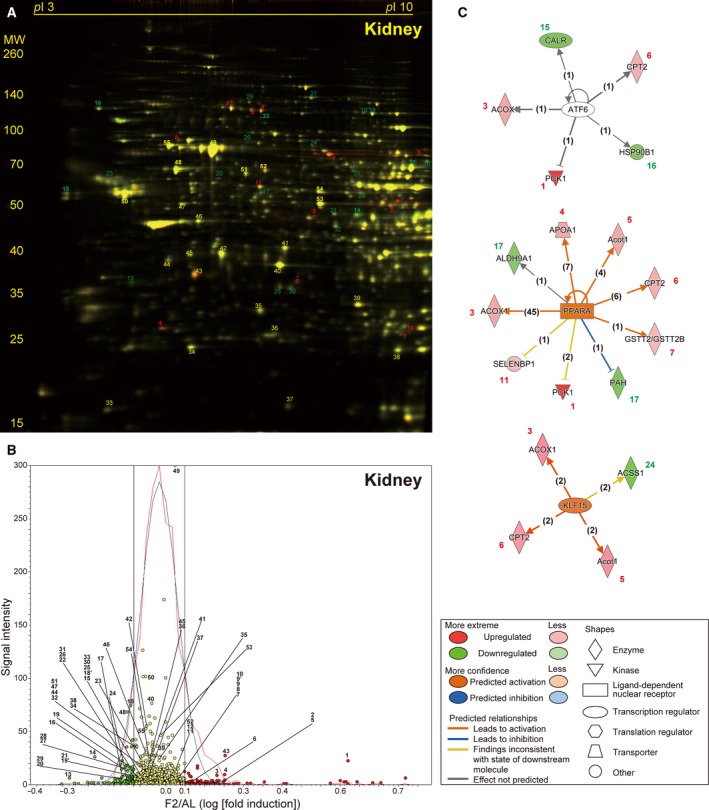
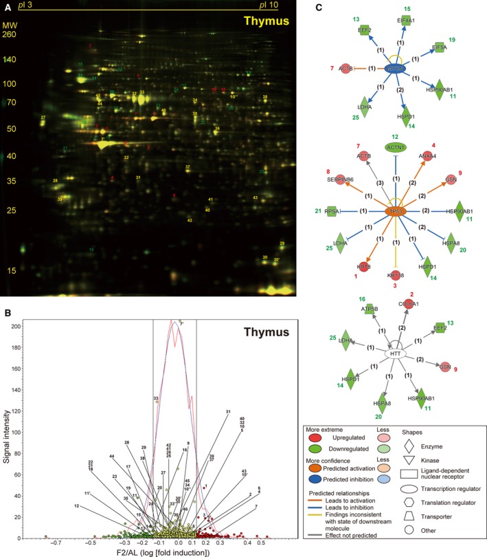
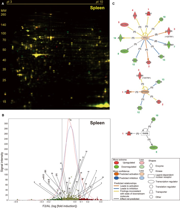
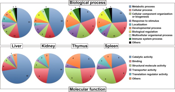
Overnight fasting is a routine procedure before surgery in clinical settings. Intermittent fasting is the most common diet/fitness trend implemented for weight loss and the treatment of lifestyle-related diseases. In either setting, the effects not directly related to parameters of interest, either beneficial or harmful, are often ignored. We previously demonstrated differential activation of cellular adaptive responses in 13 atrophied/nonatrophied organs of fasted mice by quantitative PCR analysis of gene expression. Here, we investigated 2-day fasting-induced protein remodeling in six major mouse organs (liver, kidney, thymus, spleen, brain, and testis) using two-dimensional difference gel electrophoresis (2D DIGE) proteomics as an alternative means to examine systemic adaptive responses. Quantitative analysis of protein expression followed by protein identification using matrix-assisted laser desorption ionization-time-of-flight mass spectrometry (MALDI-TOFMS) revealed that the expression levels of 72, 26, and 14 proteins were significantly up- or downregulated in the highly atrophied liver, thymus, and spleen, respectively, and the expression levels of 32 proteins were up- or downregulated in the mildly atrophied kidney. Conversely, there were no significant protein expression changes in the nonatrophied organs, brain and testis. Upstream regulator analysis highlighted transcriptional regulation by peroxisome proliferator-activated receptor alpha (PPARα) in the liver and kidney and by tumor protein/suppressor p53 (TP53) in the thymus, spleen, and liver. These results imply of the existence of both common and distinct adaptive responses between major mouse organs, which involve transcriptional regulation of specific protein expression upon short-term fasting. Our data may be valuable in understanding systemic transcriptional regulation upon fasting in experimental animals.

Keywords: 2D DIGE; PPARα; TP53; fasting; proteomics; transcriptional regulation.


Figures

Figure 1
Figure 1
Impact of 1‐ or 2‐day fasting on serum biochemical parameters. Serum levels of glucose, insulin, C‐peptide 2, leptin, resistin, ketone bodies, gastric inhibitory polypeptide (GIP), and adiponectin were measured. AL, ad libitum‐fed (AL); F1, 1‐day fasted; F2, 2‐day fasted. Data are mean ± SD (n: sample numbers); significant changes in *P < 0.05, **P < 0.01, and ***P < 0.001 vs AL; ### P < 0.001 vs F1 by one‐way ANOVA with Tukey's multiple comparison test.
Figure 2
Figure 2
Fasting‐induced protein remodeling in the liver. Fluorescent 2D DIGE was performed on liver homogenates from ad libitum‐fed (AL) and 2‐day fasted (F2) mice. (A) Representative fluorescent gel image in which proteins upregulated by fasting are labeled in red and those downregulated are in green. Approximate isoelectric points (pI ) and molecular weights (MW; kDa) are indicated. (B) Quantitative profiling of the above image using DeCyder software. Upregulated and downregulated (> 1.1‐fold) protein spots are labeled in red and green, respectively, with others in yellow. The x‐axis represents log [(Fasted/AL) fold induction], and the y‐axis represents spot signal intensity; the red line represents spot number distribution, while the blue line its Gaussian approximation; and two black straight lines represent 1.1 and –1.1 fold change. (C) The three highest scoring upstream regulators (PPARα, NR1I2, and PPARγ) listed by Ingenuity Pathway Analysis (IPA) of the samples from AL and F2 mice (n = 4 each). Upregulated proteins with IDs identical to those in A and B are shown in red and downregulated proteins are in green, and predicted relationships are indicated by various types of lines described in the panel. The numbers in parentheses are the numbers of current publications reporting those relationships.
Figure 3
Figure 3
Fasting‐induced protein remodeling in the kidney. Fluorescent 2D DIGE was performed on kidney homogenates from ad libitum‐fed (AL) and 2‐day fasted (F2) mice. (A) Representative fluorescent gel image in which proteins upregulated by fasting are labeled in red and those downregulated are in green. (B) Quantitative profiling of the above image using DeCyder. (C) The three highest scoring upstream regulators (ATF6, PPARα, and KLF15) listed by IPA of the samples from AL and F2 mice (n = 4 each). Refer to Fig. 2 legend for detailed information.
Figure 4
Figure 4
Fasting‐induced protein remodeling in the thymus. Fluorescent 2D DIGE was performed on thymus homogenates from ad libitum‐fed (AL) and 2‐day fasted (F2) mice. (A) Representative fluorescent gel image in which proteins upregulated by fasting are labeled in red and those downregulated are in green. (B) Quantitative profiling of the above image using DeCyder. (C) The three highest scoring upstream regulators (MYCN, TP53, and HTT) listed by IPA of the samples from AL and F2 mice (n = 4 each). Refer to Fig. 2 legend for detailed information.
Figure 5
Figure 5
Fasting‐induced protein remodeling in the spleen. Fluorescent 2D DIGE was performed on spleen homogenates from ad libitum‐fed (AL) and 2‐day fasted (F2) mice. (A) Representative fluorescent gel image in which proteins upregulated by fasting are labeled in red and those downregulated are in green. (B) Quantitative profiling of the above image by DeCyder. (C) The three highest scoring upstream regulators (TP53, NFKBIA, and RARB) listed by IPA of the samples from AL and F2 mice (n = 4 each). Refer to Fig. 2 legend for detailed information.
Figure 6
Figure 6
Venn diagrams for clarification of identified proteins that are involved in various biological processes and molecular functions. Fasting‐regulated proteins in the liver, kidney, thymus, and spleen are categorized by ‘biological process’ or ‘molecular function’ using PANTHER software.
Figure 7
Figure 7
Fasting‐induced protein remodeling in the brain and testis. Fluorescent 2D DIGE was performed on brain (A and B) and testis (C and D) homogenates from ad libitum‐fed (AL) and 2‐day fasted (F2) mice. (A and C) Representative fluorescent images, in which proteins upregulated by fasting are labeled in red and those downregulated are in green. (B and D) Quantitative profiling of the above images using DeCyder software. Refer to Fig. 2 legend for detailed information.

Similar articles

Cited by

References

    1. Longo VD and Mattson MP (2014) Fasting: molecular mechanisms and clinical applications. Cell Metab 19, 181–192. - PMC - PubMed
    1. van Praag H, Fleshner M, Schwartz MW and Mattson MP (2014) Exercise, energy intake, glucose homeostasis, and the brain. J Neurosci 34, 15139–15149. - PMC - PubMed
    1. Jensen TL, Kiersgaard MK, Sorensen DB and Mikkelsen LF (2013) Fasting of mice: a review. Lab Anim 47, 225–240. - PubMed
    1. Kersten S, Seydoux J, Peters JM, Gonzalez FJ, Desvergne B and Wahli W (1999) Peroxisome proliferator‐activated receptor alpha mediates the adaptive response to fasting. J Clin Invest 103, 1489–1498. - PMC - PubMed
    1. Leone TC, Weinheimer CJ and Kelly DP (1999) A critical role for the peroxisome proliferator‐activated receptor alpha (PPARalpha) in the cellular fasting response: the PPARalpha‐null mouse as a model of fatty acid oxidation disorders. Proc Natl Acad Sci USA 96, 7473–7478. - PMC - PubMed

LinkOut - more resources