. 2018 Aug 29;4(8):e00753.
doi: 10.1016/j.heliyon.2018.e00753. eCollection 2018 Aug.

Food-derived polyphenols inhibit the growth of ovarian cancer cells irrespective of their ability to induce antioxidant responses

Affiliations

Food-derived polyphenols inhibit the growth of ovarian cancer cells irrespective of their ability to induce antioxidant responses

Youngjoo Kwon. Heliyon. .

Abstract

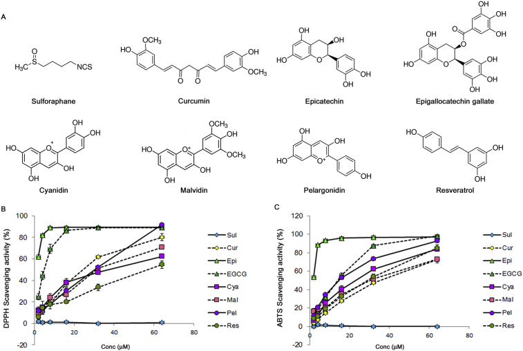
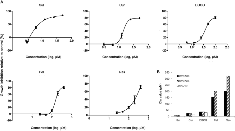
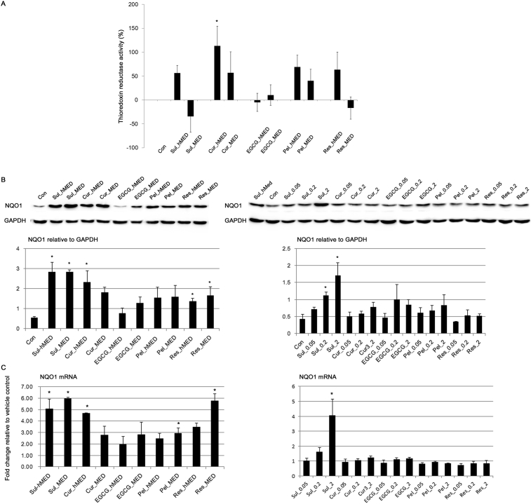
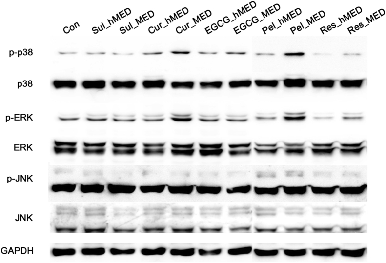
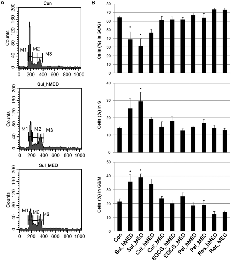
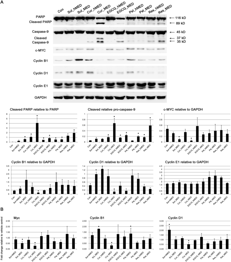
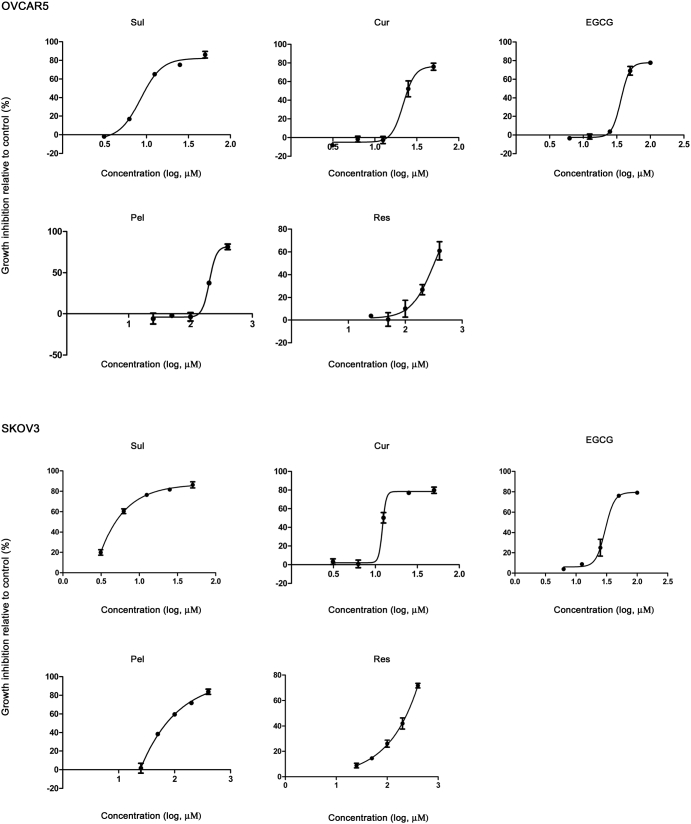
The use of plant polyphenols to prevent cancer has been studied extensively. However, recent findings regarding the cancer-promoting effects of some antioxidants have led to reservations regarding the therapeutic use of food-derived antioxidants including polyphenols. The aim of this study was to evaluate the therapeutic potential of food-derived polyphenols and their use and safety in cancer patients. The free-radical scavenging ability of sulforaphane and various food-derived polyphenols including curcumin, epigallocatechin gallate, epicatechin, pelargonidin, and resveratrol was compared with their growth inhibitory effect on ovarian cancer cells. Oxidative stress and/or antioxidant responses and anti-proliferative pathways were evaluated after administering sulforaphane and polyphenols at doses at which they have been shown to inhibit the growth of ovarian cancer cells. No correlation was observed between their ability to scavenge free radicals and their ability to inhibit the growth of ovarian cancer cells. With the exception of epigallocatechin gallate, all of the antioxidants that were tested at doses that inhibited cell growth significantly increased NAD(P)H quinone dehydrogenase I (NQO1) expression but induced cell cycle arrest and/or apoptotic signaling. Epigallocatechin gallate exhibited a higher free radical scavenging activity but did not induce NQO1 expression at either the mRNA or at the protein level. Treatment with polyphenols at physiological doses did not significantly alter the growth of ovarian cancer cells or NQO1 expression. Therefore, individual food-derived polyphenols appear to have different anti-cancer mechanisms. Their modes of action in relation to their chemical properties should be established, rather than collectively avoiding the use of these agents as antioxidants.

Keywords: Biochemistry; Cancer research; Cell biology; Food science.


Figures

Fig. 1
Fig. 1
Comparison of free-radical scavenging by various food-derived polyphenols. (A) The chemical structures of sulforaphane and the various food-derived polyphenols that were tested in this study. The ability to scavenge DPPH radicals (B) and ABTS radical cation (C) was measured at final polyphenol concentrations ranging from 0 to 64 μM.
Fig. 2
Fig. 2
Inhibition of the growth of ovarian cancer cells by treatment with various food-derived polyphenols and sulforaphane. Ovarian cancer cells were cultured overnight in 96-well plates and then treated with 0–400 μM polyphenols and sulforaphane for 72 h. (A) Cell-growth inhibition relative to the vehicle control is shown in OVCAR3 cells. (B) The IC50 (half-maximal inhibitory concentration) values for OVCAR3, OVCAR5, and SKOV3 cells.
Fig. 3
Fig. 3
The effect of food-derived polyphenols on endogenous antioxidant enzymes. (A) Thioredoxin reductase activity was measured 24 h after treatment of OVCAR3 with food-derived polyphenols and sulforaphane at their maximal effective dose (MED) and half of MED (hMED). (B) The effect of food polyphenols and sulforaphane on the protein expression level of NQO1 was assessed by immunoblotting after treatment at the MED and hMED (left panel) and 20, 50 nM, and 2 μM of polyphenols (right panel) for 24 h. The results of the densitometric analysis of the blots are shown in the lower panel. Full, non-adjusted blots are available in Supplementary Fig. 2. (C) The NQO1 mRNA level was measured by RT-qPCR. Values are the mean ± SE, n = 3. An asterisk (*) represents a significant difference (p < 0.05) compared to the vehicle control.
Fig. 4
Fig. 4
Activation of mitogen-activated protein kinases (MAPKs) by food-derived polyphenols. Activation of MAPKs [p38; extracellular signal-regulated kinase (ERK); c-Jun NH2-terminal kinase (JNK)] in OVCAR3 cells was assessed by immunoblotting after a 6 h treatment at the maximal effective dose (MED) and half of MED (hMED) of the polyphenols and sulforaphane. The data are representative of three independent experiments. GAPDH, glyceraldehyde-3-phosphate dehydrogenase. Full, non-adjusted blots are available in Supplementary Fig. 2.
Fig. 5
Fig. 5
The effect of food-derived polyphenols on cell cycle progression. OVCAR3 cells were treated with polyphenols and sulforaphane at their maximal effective doses (MED) and half of MED (hMED) and the DNA content of the cells was analyzed by flow cytometry. (A) Representative flow cytometry plots. (B) The percentage of cells in each phase of the cell cycle. Values are the mean ± SE, n = 3. An asterisk (*) represents a significant difference (p < 0.05) compared to the vehicle control. M1, G0/G1 phase; M2, S phase; M3, G2/M phase.
Fig. 6
Fig. 6
Induction of anti-proliferative pathways by food-derived polyphenols. (A) The effect of food polyphenols on protein expression levels was assessed by immunoblotting after 24 h of treatment at the MED and hMED of the polyphenols and sulforaphane. The results for the densitometric analysis of blots are shown in the lower panel. PARP, poly (ADP-ribose) polymerase (PARP); GAPDH, glyceraldehyde-3-phosphate dehydrogenase. Full, non-adjusted blots are available in Supplementary Fig. 2. (B) Transcription levels of the indicated genes were measured by RT-qPCR. Values are the mean ± SE, n = 3. An asterisk (*) represents a significant difference (p < 0.05) compared to the vehicle control.
Fig. 7
Fig. 7
The effect of food-derived polyphenols on the levels of cytokines secreted by fibroblasts. Serum-starved fibroblasts were treated with polyphenols and sulforaphane (3.5 μM), and changes in the levels of cytokines secreted by the fibroblasts were measured. Values are the mean ± SE, n = 3. An asterisk (*) represents a significant difference (p < 0.05) compared to the vehicle control.
Supplementary Figure 1
Inhibition of growth of ovarian cancer cells by treatment with various food-derived polyphenols and sulforaphane. Cell-growth inhibition relative to the vehicle control is shown in OVCAR5 and SKOV3 cells.

Similar articles

Cited by

References

    1. Key T.J., Schatzkin A., Willett W.C., Allen N.E., Spencer E.A., Travis R.C. Diet, nutrition and the prevention of cancer. Publ. Health Nutr. 2004;7(1A):187–200. - PubMed
    1. Collins A.R. Antioxidant intervention as a route to cancer prevention. Eur. J. Cancer. 2005;41(13):1923–1930. - PubMed
    1. Sayin V.I., Ibrahim M.X., Larsson E., Nilsson J.A., Lindahl P., Bergo M.O. Antioxidants accelerate lung cancer progression in mice. Sci. Transl. Med. 2014;6(221):221ra15. - PubMed
    1. Le Gal K., Ibrahim M.X., Wiel C., Sayin V.I., Akula M.K., Karlsson C. Antioxidants can increase melanoma metastasis in mice. Sci. Transl. Med. 2015;7(308):308re8. - PubMed
    1. Tanvetyanon T., Bepler G. Beta-carotene in multivitamins and the possible risk of lung cancer among smokers versus former smokers: a meta-analysis and evaluation of national brands. Cancer. 2008;113(1):150–157. - PubMed

LinkOut - more resources