. 2018 Oct 15;47(20):7539-7551.
doi: 10.1039/c7cs00735c.

Peptide supramolecular materials for therapeutics

Affiliations

Peptide supramolecular materials for therapeutics

Kohei Sato et al. Chem Soc Rev. .

Abstract

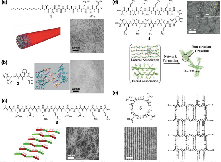
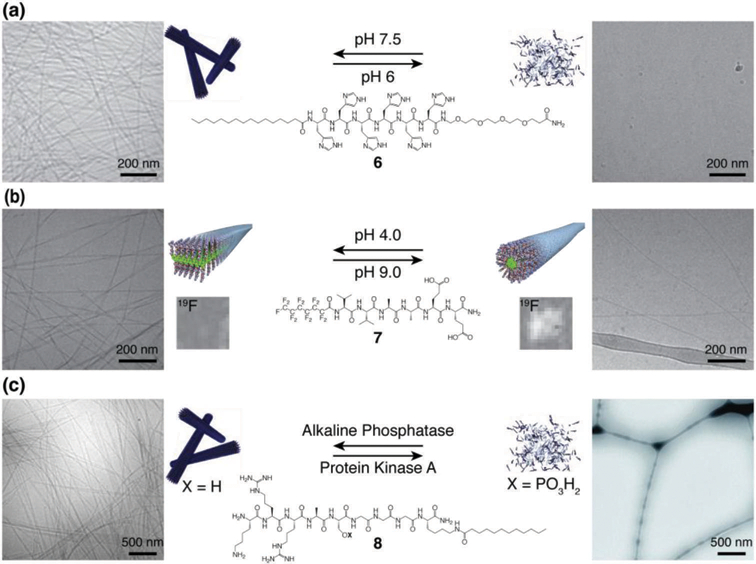
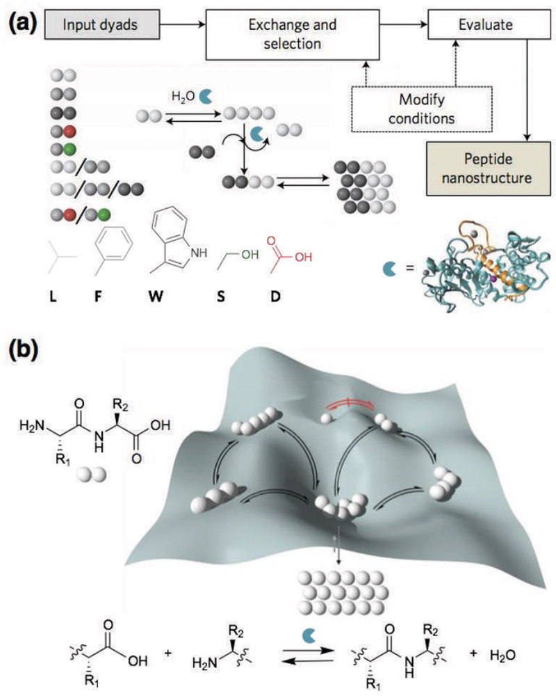
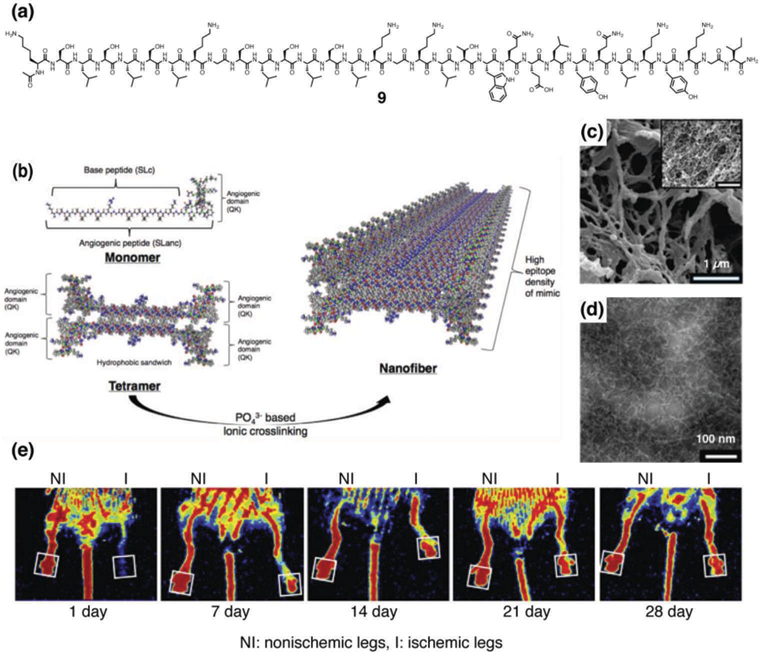
Supramolecular assembly of peptide-based monomers into nanostructures offers many promising applications in advanced therapies. In this Tutorial Review, we introduce molecular designs to control the structure and potential biological function of supramolecular assemblies. An emphasis is placed on peptide-based supramolecular nanostructures that are intentionally designed to signal cells, either directly through the incorporation of amino acid sequences that activate receptors or indirectly by recruiting native signals such as growth factors. Additionally, we describe the use and future potential of hierarchical structures, such as single molecules that assemble into nanoscale fibers which then align to form macroscopic strings; the strings can then serve as scaffolds for cell growth, proliferation, and differentiation.


Figures

Fig. 1
Fig. 1
(a) Chemical structure of a peptide amphiphile (PA), schematic illustration of its supramolecular assembly into a cylindrical nanofiber, and cryogenic transmission electron microscopy (cryo-TEM) of nanofibers. Reprinted with permission from ref. 32 (Copyright 2015 Wiley-VCH). (b) Chemical structure of an Fmoc-dipeptide, schematic illustration of molecular packing (Fmoc groups: orange, phenyl groups: purple), and cryogenic scanning electron microscopy (cryo-SEM) of nanofibers. Reprinted with permission from ref. 15 (Copyright 2008 Wiley-VCH). (c) Chemical structure of the Z-DNA-binding mimetic amphipathic peptide, schematic illustration of its supramolecular assembly, and SEM of nanofibers. Reprinted with permission from ref. 16 (Copyright 1993 National Academy of Sciences). (d) Chemical structure of a β-hairpin peptide, schematic illustration of its folding and supramolecular assembly, and TEM of nanofibers. Reprinted with permission from ref. 19 (Copyright 2007 National Academy of Sciences). (e) Chemical structure of a cyclic peptide with alternating d- and l-amino acids and its tubular assembly, and TEM of closely packed nanotubes. Reprinted with permission from ref. 21 (Copyright 1993 Nature Publishing Group).
Fig. 2
Fig. 2
Structures of reversibly responsive PAs with schematic illustrations and cryogenic transmission electronic micrographs of their supramolecular assemblies. (a) pH-responsive histidine-based PA 6 at pH 7.5 (left) and pH 6.0 (right), showing the assembled fibers and dissembled states, respectively. (b) pH-responsive PA 7 with a perfluorinated tail at pH 4.0 (left) and pH 9.0 (right), showing the assembled ribbons and fibers, respectively. F MRI solution images of the assemblies are shown above the schematic illustrations. (c) Enzyme-responsive PA 8 in the de-phosphorylated form after reaction with alkaline phosphatase (left) and phosphorylated form (right) after conversion with protein kinase A, showing the assembled fibers and dissembled states, respectively. Adapted with permission from ref. 11, ref. 12, and ref. 13 (Copyright (a, b) 2014 American Chemical Society, (c) 2011 Royal Society of Chemistry).
Fig. 3
Fig. 3
(a) Schematic illustration of the dynamic peptide library system to search for supramolecular materials, which involves mixtures of dipeptides (input dyads), dynamic exchange of peptide sequences catalyzed by enzymatic condensation, hydrolysis, and transacylation, with the most-stable assembling structure eventually emerging (peptide nanostructure). The reaction conditions may be modified to promote or reduce certain supramolecular interactions and thermodynamic selection. (b) Potential energy surface that shows the formation of peptide oligomers. The depth of the wells represents the relative stability of the assembling peptides formed. Reprinted with permission from ref. 22 (Copyright 2016 Nature Publishing Group).
Fig. 4
Fig. 4
(a) Chemical structure of a vascular endothelial growth factor (VEGF)-mimetic multi-domain peptide 9. (b) Schematic illustrations of supramolecular assembly of 9 to form nanofibers. (c) SEM image of nanofibers formed by 9. (d) TEM of nanofibers formed by 9. (e) Recovery from hind limb ischemia after treatment with VEGF-mimetic multidomain peptide 9. Laser Doppler perfusion imaging showed rapid restoration of blood flow to the foot pad (boxed region) in VEGF-mimetic multidomain peptide-treated 13-month old mice. Reprinted with permission from ref. 24 and ref. 25 (Copyright 2015 American Chemical Society, 2016 Elsevier).
Fig. 5
Fig. 5
(a) Chemical structures of heparin and a heparin-mimetic PA 10. (b) SEM image of nanofibers formed by 10. (c) TEM of nanofibers formed by 10. (d) In vitro angiogenesis assay. Bright field image of HUVECs cultured on nanofiber matrix formed by PA 10 (100x magnification). (e–g) Evaluation of in vivo bioactivity by corneal angiogenesis assay. (e) Injection of hydrogel formed by 1wt% of PA 2 with 10 ng of VEGF and bFGF-induced vascularization in cornea. (f) Application of growth factor solution (10 ng of VEGF and bFGF) in physiological saline (without PA gel) did not induce vascularization. (g) Ratio of vascularized area to total area was calculated for both groups. (h–i) Representative µCT images of bones treated with (h) PA 10 hydrogel and (i) saline solution after 4 weeks. (j–l) Histological evaluation of a tibial defect model treated with (j) PA 10 hydrogel and (k) saline solution after 4 weeks stained with H&E. Scale bars are 200 µm. (l) Regenerated bone areas were quantified through the histological evaluation of H&E results. Reprinted with permission from ref. 27 and ref. 30 (Copyright 2011 American Chemical Society, 2016 Royal Society of Chemistry).
Fig. 6
Fig. 6
(a) Chemical structures of glycol-PAs 1114 and backbone PA 15 lacking the glycol unit. (b) Cryogenic TEM image and a schematic illustration of PA 13 to form nanofibers. (c) Plot of alkaline phosphatase (ALP) activity in C2C12 cells treated with BMP-2 and glycol-PAs 1114 as a function of increasing monosaccharide density on the nanostructures (treatment with heparin is indicated by the dashed line). (d) Quantification of Western blots of C2C12 cells stimulated for 3 h with BMP-2 revealing the effect of heparin or PA 11 nanofibers on Smad phosphorylation. Cells were also treated with LDN to inhibit BMP-2 signaling. (e) Representative volume renderings from µCT (yellow arrows indicate fusion). (f) Digital sagittal section through the fusion mass from an animal treated with 100 ng BMP-2 and PA 11 nanostructures. (g) Representative sagittal cross-sectional images of L4–L5 posterolateral spine specimens with H&E staining. Reprinted with permission from ref. 33 (Copyright 2017 Nature Publishing Group).
Fig. 7
Fig. 7
(a) Chemical structures of RGDS epitope-presenting PA 16 in which a glycine linker of variable length (n = 1, 3, and 5) presents the epitope, and base PA 17 lacking the RGDS sequence. (b–d) Cryogenic TEM images and schematic illustrations of RGDS epitope-presenting PAs co-assembled with base PA 17 at a 1:9 weight ratio. (b) PA 16 (n = 1), (c) PA 16 (n = 3) and (d) PA 16 (n = 5). (e) Representative images of 3T3 fibroblasts cultured on PA-coated substrates for 5 h and stained with phalloidin for actin filaments. (f) Cell morphology on PA substrates, compared by measuring the projected cell area. Reprinted with permission from ref. 43 (Copyright 2015 Royal Society of Chemistry).
Fig. 8
Fig. 8
(a, b) SEM images of aligned PA fibers within macroscopic strings made by dragging annealed PA solutions into a calcium chloride solution. (c) A PA solution dyed with trypan blue extruded into phosphate-buffered saline solution after annealing. (d) Transmission electron micrograph of precipitated collagen-like PA. (e) AFM image (top) and linecut (bottom) of periodic microstructure of collagen-like PA, showing a periodic, undulating pattern. (f) Photos of mixtures of collagen-like PA and non-collagen-like control PA (1 wt% on DI water), ranging from pure collagen-like PA (1, 8) to pure control PA (2, 3). Reprinted with permission from ref. 45 (a-c) and ref. 49 (d-f) (Copyright 2010 Nature Publishing Group, 2015 American Chemical Society).

References

    1. Aida T, Meijer EW and Stupp SI, Science, 2012, 335, 813–817. - PMC - PubMed
    1. Tabony J, Biol. Cell, 2006, 98, 603–617. - PubMed
    1. Hamley IW, Angew. Chem. Int. Ed. Engl, 2014, 53, 6866–6881. - PubMed
    1. Berndt P, Fields GB and Tirrell M, J Am Chem Soc, 1995, 117, 9515–9522.
    1. Hartgerink JD, Beniash E and Stupp SI, Science, 2001, 294, 1684–1688. - PubMed

MeSH terms

Substances