

Breast implant-associated anaplastic large cell lymphoma: a pictorial review
- PMID: 30187266
- PMCID: PMC6206369
- DOI: 10.1007/s13244-018-0652-z
Breast implant-associated anaplastic large cell lymphoma: a pictorial review
Abstract
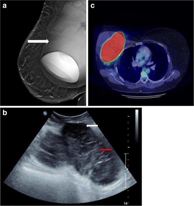
Breast implant-associated anaplastic large cell lymphoma (BIA-ALCL) is a newly described and rare T-cell lymphoma of the breast. Since 2007, there have been 56 cases of confirmed BIA-ALCL in Australia and New Zealand. The incidence is believed to be on the rise as the prevalence of elective breast implantation increases. In 2016, the World Health Organization (WHO) classified BIA-ALCL as a recognised entity and emphasised the importance of surgical management of the disease. BIA-ALCL typically presents as a delayed, non-infective fluid collection around a textured breast implant or residual fibrous scar capsule. The mean age of presentation is 47 years, with an average time frame of 7.5 years following breast implantation. Although rare, BIA-ALCL is increasing in incidence. To avoid delays in diagnosis, radiologists should consider this form of lymphoma in the differential of any non-acute peri- or post-prosthetic effusion, and suggest cytological evaluation, so as not to miss this rare but important diagnosis. TEACHING POINTS: • BIA-ALCL is a newly described and rare T-cell lymphoma of the breast. • Since 2007, there have been 56 cases of confirmed BIA-ALCL in Australia and New Zealand. • BIA-ALCL presents as a delayed, non-infective fluid collection. • The effusion typically accumulates around a textured breast implant or residual fibrous capsule.
Keywords: Breast imaging; Lymph; Nuclear imaging; Oncologic imaging; Ultrasound.
Figures
References
-
- Loch-Wilkinson A, Beath KJ, Knight RJW, Wessels WLF, Magnusson M, Papadopoulos T, et al. Breast implant-associated anaplastic large cell lymphoma in Australia and New Zealand: high-surface-area textured implants are associated with increased risk. Plast Reconstr Surg. 2017;140(4):645–654. doi: 10.1097/PRS.0000000000003654. - DOI - PubMed
-
- Australian Government (2018) Breast implants and anaplastic large cell lymphoma. Therapeutic Goods Administration, Department of Health, Australian Government. Available online at: https://www.tga.gov.au/node/733565
-
- Binmahfouz A, Steinke K. A case report of breast implant-associated anaplastic large cell lymphoma: the good, the bad, and the ugly. Int J Case Rep Images. 2016;7(8):537–541.
-
- Swerdlow S. H., Campo E., Pileri S. A., Harris N. L., Stein H., Siebert R., Advani R., Ghielmini M., Salles G. A., Zelenetz A. D., Jaffe E. S. The 2016 revision of the World Health Organization classification of lymphoid neoplasms. Blood. 2016;127(20):2375–2390. doi: 10.1182/blood-2016-01-643569. - DOI - PMC - PubMed
Publication types
LinkOut - more resources
Full Text Sources
Other Literature Sources
