

Case Reports
doi: 10.1111/jmp.12373.
Epub 2018 Sep 6.
Eosinophilic aortitis with thoracic aortic aneurysm and rupture in a captive-born owl monkey
Affiliations
- PMID: 30187922
- PMCID: PMC11025315
- DOI: 10.1111/jmp.12373
Item in Clipboard
Case Reports
Eosinophilic aortitis with thoracic aortic aneurysm and rupture in a captive-born owl monkey
J Med Primatol.
2018 Dec.
Abstract
Eosinophilic aortitis is a rare condition in animals and humans, and it has been occasionally reported associated with parasitic migration and with a poorly understood complex group of autoimmune vasculitides. Here, we describe a case of eosinophilic aortitis with thoracic aortic aneurysm and rupture in a captive-born owl monkey and discuss the differential diagnoses.
Keywords: Aotus nancymai; aorta; arteritis; autoimmune; eosinophil; granulomatosis; nonhuman primate; vasculitides.
Published 2018. This article is a U.S. Government work and is in the public domain in the USA.
Figures
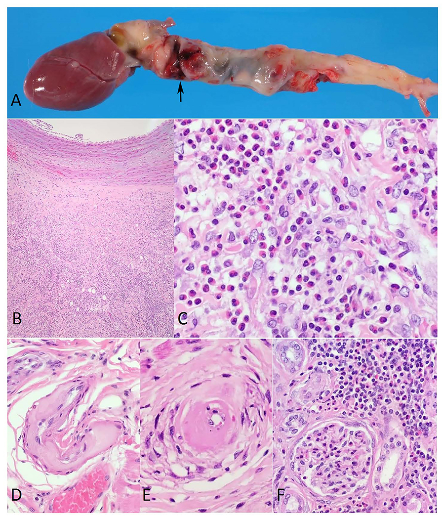
A. Owl monkey, aorta. Photograph showing entire length of the thoracic aorta out of the heart to the aortic hiatus in the diaphragm dilated and irregularly shaped with an approximately 5 mm diameter tear (arrow). B. Owl monkey, aorta. Low power micrograph of the thoracic aorta wall showing marked intimal thickening due to accumulation of mucinous ground substance, loss of the elastic lamina, separation of the tunica media by a space filled with blood and an outer band composed of abundant granulation tissue, fibroplasia, edema, mucinous deposits, and moderate infiltrate of eosinophils. H&E stain. 40x original magnification. C. Owl monkey, aorta. Micrograph showing at higher magnification the affected aorta infiltrated with eosinophils. H&E stain. 400x original magnification. D. Owl monkey, heart. Micrograph of a medium-sized coronary artery with extensive fibrinoid necrosis and hyaline change affecting most of the vessel wall. H&E stain. 200x original magnification. E. Owl monkey, heart. Micrograph of a small coronary artery with marked hyaline change surrounded by vacuolated and necrotic vascular myocytes. H&E stain. 400x original magnification. F. Owl monkey, kidney. Micrograph showing multifocal mild to moderate interstitial lymphoeosinophilic infiltrates, foci of interstitial and periglomerular fibrosis, degenerate cortical tubules, and tubules with attenuated epithelium filled with eosinophilic granular cast material. H&E stain. 200x original magnification.
References
-
- Albert DL, Brodie SJ, Sasseville VG, Ringler DJ. Peripheral blood eosinophilia in owl monkeys is associated with increased eosinophilopoeisis yet depressed recruitment kinetics to the Chemokine RANTES. Blood 1996; 88(5):1718–1724. - PubMed
-
- Animal Welfare Act as Amended. 2013. 7 USC § 2131-2159.
-
- Animal Welfare Regulations. 2013. 9 CFR § 3.75-3.92.
-
- Baer JF, Gibson SV, Weller RE, Buschbom RL, Leathers CW. Naturally occurring aortic aneurysms in owl monkeys (Aotus spp.). Lab Anim Sci 1992; 42(5):463–466. - PubMed
Publication types
MeSH terms
Grants and funding
LinkOut - more resources
Full Text Sources
Other Literature Sources
