. 2018 Sep 6;12(9):e0006738.
doi: 10.1371/journal.pntd.0006738. eCollection 2018 Sep.

Phylogenomic analysis unravels evolution of yellow fever virus within hosts

Affiliations

Phylogenomic analysis unravels evolution of yellow fever virus within hosts

Chen Chen et al. PLoS Negl Trop Dis. .

Abstract

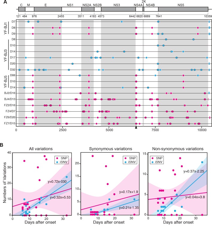
The yellow fever virus (YFV) recently reemerged in the large outbreaks in Africa and Brazil, and the first imported patients into Asia have recalled the concerns of YFV evolution. Here we show phylogenomics of YFV with serial clinical samples of the 2016 YFV infections. Phylogenetics exhibited that the 2016 strains were close to Angola 1971 strains and only three amino acid changes presented new to other lineages. Deep sequencing of viral genomes discovered 101 intrahost single nucleotide variations (iSNVs) and 234 single nucleotide polymorphisms (SNPs). Analysis of iSNV distribution and mutated allele frequency revealed that the coding regions were under purifying selection. Comparison of the evolutionary rates estimated by iSNV and SNP showed that the intrahost rate was ~2.25 times higher than the epidemic rate, and both rates were higher than the long-term YFV substitution rate, as expected. In addition, the result also hinted that short viremia duration of YFV might further hinder the evolution of YFV.


Conflict of interest statement

The authors have declared that no competing interests exist.

Figures

Fig 1
Fig 1. Cases of twelve documented returning YFV patients in China by date in the outbreak of Angola from March to May 2016.
Timeline of events for each patient from symptom onset to the leaving from hospital are shown with different molecular detection methods and results. Dots denote that we did not get any positive molecular detection result in the time point, grey (before admitted to hospital) and blue (after admitted to hospital). The larger size dots means that we performed Real-time PCR detections in that point. The red triangles denote the sample have been detected by PCR and Real-time PCR with positive results. Blue boxes denote the samples that were subjected to NGS analysis at the time points.
Fig 2
Fig 2. Phylogenetics and amino acids comparison of YFV viruses.
(A) Maximum-likelihood phylogenetic tree of YFV genome sequences (the first 130 bps in 5’-UTR and the last 370 bps in 3’-UTR sequences are excluded). Bootstrap support values are shown along the branches. Wesselsbron virus, Sepik virus and Entebbe bat virus were used as outgroup. (B) Maximum-likelihood phylogenic tree of YFV genomes of Angola 2016 strains. Viruses from patients with severe disease are highlighted in magenta. Angola 1971(GenBank accession AY968064) was used as outgroup. Bootstrap support values (≥70) are shown. (C) Comparison of amino acids of YFV consensus sequence of each lineage in (A). Identical amino acid to vaccine strain (17D) lineage is denoted as a dot. The amino acid sites that were different in Angola 2016 and 1971, are highlight with red rectangles. Novel amino acid substitutions of Angola 2016 are denoted by arrowheads.
Fig 3
Fig 3. The iSNVs of Angola 2016 YFVs from clinical samples.
(A) Sequencing depth across the sequenced genomes. The x-axis represents the YFV genome, with the ORF boundaries indicated by vertical dashed lines. The sequencing depth smoothed by locally weighted smooth regression (LOESS) is shown by the black curve, with 95% confidence interval as shown by shadow. (B) Numbers of iSNVs at non-coding (NC) regions and codon positions of the ORF. (C) Numbers of nonsynonymous (N) and synonymous (S) iSNVs of the ORF. (D) Box plots of the mutated allele frequencies (MuAFs) for non-coding, synonymous and nonsynonymous iSNVs. The MuAF values of three kinds of iSNVs are compared to each other with the Wilcoxon rank-sum test. Dashed lines denote the boundaries of MuAFs in iSNV identification. Boxes denote the interquartile range (IQR) between the first and third quartiles. Lines inside the boxes indicate the median, and the lines outside boxes represent values within 1.5 times the IRQ. Outliers are denoted as dots. (E) Comparison of MuAF spectra of non-coding, synonymous and nonsynonymous iSNVs. MuAFs (dots) are shown with LOESS lines (95% confidence interval in shadow). Expectation under neutral selection is shown by a black line. (F) Prediction of RNA secondary structures of 3’ UTR (with 68 bp downstream of the stop codon). The earliest type of Angola 2016 (wildtype) and two nucleotides variant types are illustrated. Six variation sites are highlighted in magenta. The stop codon is in the gray box.
Fig 4
Fig 4. Genomic variations of Angola 2016 YFV and evolutionary model.
(A) SNPs (in magenta) and iSNVs (in cyan) detected in Angola 2016 strains along the YFV genome. Non-synonymous variations are denoted by ovals, synonymous variations by diamonds, and variations in UTR by rectangles. Non-synonymous site at 6463 is denoted by arrowhead. YFV genome of patient YF-BJ1 at 7 days after the onset (YF-BJ1/7D) was used as the reference sequence (B) Linear regression of number of variations at all variations, synonymous and non-synonymous sites accumulated by days after onset. The smooth shadows denote the LOESS fit with 95% confidence interval.

Similar articles

Cited by

References

    1. Visser LG, Roukens AH. Modelling a way out of yellow fever. Lancet 2016; 388:2847–8. 10.1016/S0140-6736(16)31330-7 - DOI - PubMed
    1. Bryant JE, Holmes EC, Barrett AD. Out of Africa: a molecular perspective on the introduction of yellow fever virus into the Americas. PLoS pathogens 2007; 3:e75 10.1371/journal.ppat.0030075 - DOI - PMC - PubMed
    1. Mutebi J-P, Rijnbrand RC, Wang H, et al. Genetic relationships and evolution of genotypes of yellow fever virus and other members of the yellow fever virus group within the Flavivirus genus based on the 3′ noncoding region. Journal of virology 2004; 78:9652–65. 10.1128/JVI.78.18.9652-9665.2004 - DOI - PMC - PubMed
    1. Stock NK, Laraway H, Faye O, Diallo M, Niedrig M, Sall AA. Biological and phylogenetic characteristics of yellow fever virus lineages from West Africa. Journal of virology 2013; 87:2895–907. 10.1128/JVI.01116-12 - DOI - PMC - PubMed
    1. Vasconcelos PF, Bryant JE, da Rosa APT, Tesh RB, Rodrigues SG, Barrett AD. Genetic divergence and dispersal of yellow fever virus, Brazil. Emerging infectious diseases 2004; 10:1578 10.3201/eid1009.040197 - DOI - PMC - PubMed

Publication types

LinkOut - more resources