. 2018 Sep 6;13(9):e0203404.
doi: 10.1371/journal.pone.0203404. eCollection 2018.

SMAD1/5 signaling in osteoclasts regulates bone formation via coupling factors

Affiliations

SMAD1/5 signaling in osteoclasts regulates bone formation via coupling factors

Amy Tasca et al. PLoS One. .

Abstract

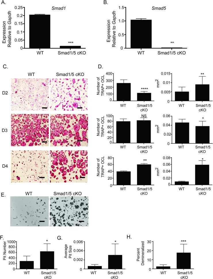
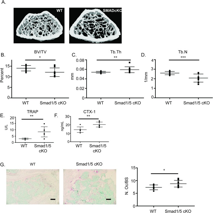
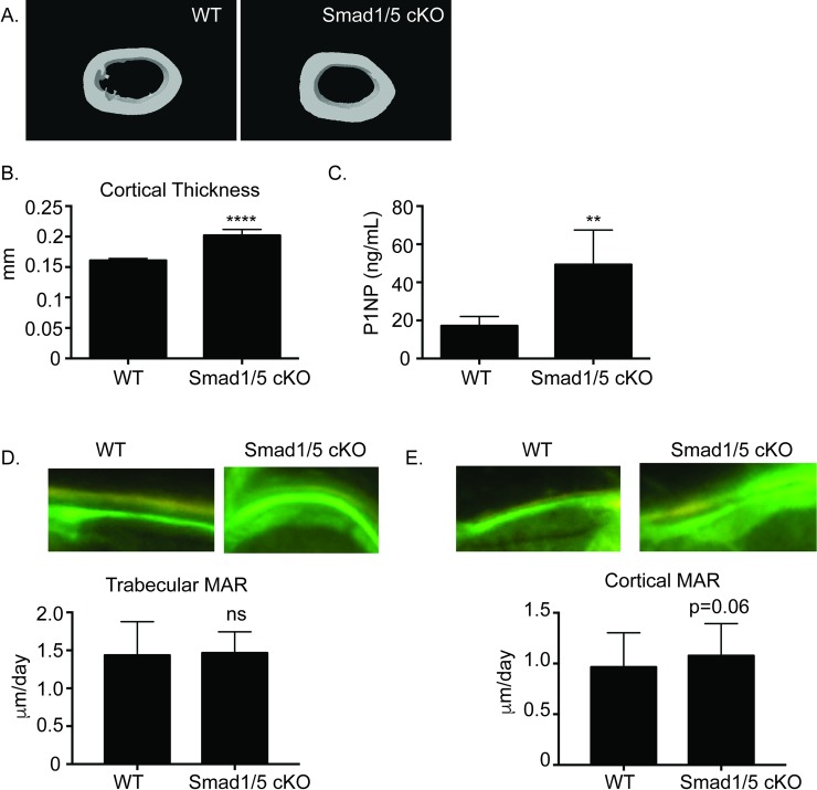
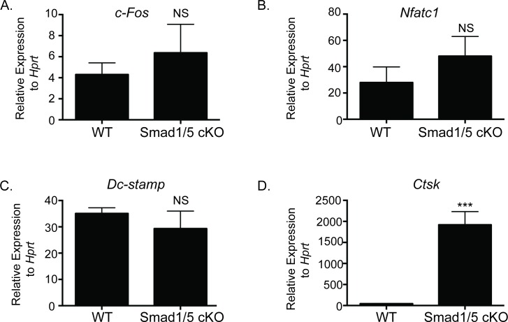
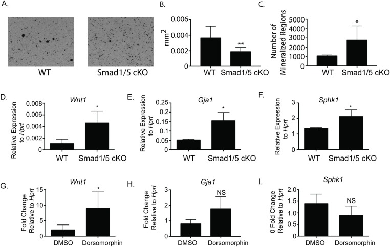
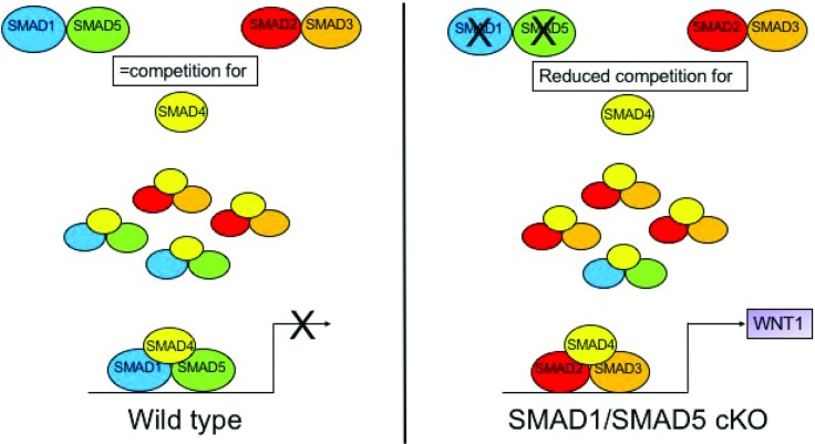
Bone remodeling occurs via coupling between bone resorption by osteoclasts and bone formation by osteoblasts. The mechanisms that regulate osteoclast signals to osteoblasts are not well understood. Published studies have reported that BMP signaling in osteoclasts regulate osteoclast coupling targets. To investigate the necessity of canonical BMP signaling on osteoclast differentiation and coupling, we mated Smad1fl/fl; Smad5fl/fl mice to c-Fms-Cre mice. We analyzed male mice at 3 months of age to determine the skeletal phenotype of the Smad1fl/fl; Smad5fl/fl;c-Fms-Cre (SMAD1/5 cKO) mice. There was a 1.2-fold decrease in trabecular BV/TV in SMAD1/5 cKO. Analyses of osteoclast serum markers in SMAD1/5 cKO mice, showed a significant increase in CTX-1 (1.5 fold) and TRAP ELISA (3 fold) compared to control mice. In these same mice, there was a 1.3-fold increase in cortical thickness. Consistent with the increase in cortical thickness, we found a 3-fold increase in osteoblast activity as measured by P1NIP ELISA assay from SMAD1/5 cKO mice. To explain the changes in cortical thickness and P1NP activity, we determined conditioned media from SMAD1/5 cKO osteoclast cultures enhanced mineralization of an osteoblast cell line and coupling factors expressed by osteoclasts that regulate osteoblast activity Wnt1 (4.5-fold increase), Gja1 (3-fold increase) and Sphk1 (1.5-fold increase) were all upregulated in osteoclasts from SMAD1/5 cKO compared to control osteoclasts. Lastly osteoclasts treated with dorsomorphin, a chemical inhibitor of SMAD1/5 signaling, demonstrates an increase in Wnt1 and Gja1 expression similar to the SMAD1/5 cKO mice. Previous studies demonstrated that TGF-β signaling in osteoclasts leads to increases in WNT1 expression by osteoclasts. Therefore, our data suggest that TGF-β and BMP signaling pathways in osteoclasts could act in an antagonistic fashion to regulate osteoblast activity through WNT1 and other coupling factors.


Conflict of interest statement

The authors have declared that no competing interests exist.

Figures

Fig 1
Fig 1. SMAD1/5 cKO mice have enhanced osteoclast differentiation and function.
BMMs were flushed from WT or SMAD1/5 cKO mice. BMMs were stimulated with M-CSF and RANKL for indicated days. (A) qRT-PCR was used to measure Smad1 gene expression following 2 days of RANKL stimulation (B) qRT-PCR was used to measure Smad5 gene expression following 2 days of RANKL stimulation (C) TRAP stained images of BMMs differentiated with M-CSF and RANKL for 2, 3, or 4 days (D) Quantification of TRAP stained images measuring number and size of TRAP positive osteoclasts. (E) Demineralization activity of WT and SMAD1/5 cKO osteoclast cultures grown on calcium phosphate surfaces. We quantified (F) pit number (G) average site of the pit (H) percent area demineralized. Scale bar 200 μm. Samples were compared using T-test* p<0.05 vs. WT, ** p<0.01 vs. WT,*** p<0.001 vs. WT, **** p<0.0001 vs. WT.
Fig 2
Fig 2. Micro CT analysis of SMAD1/5 cKO mice shows minimal change in trabecular bone.
Three-month-old male Smad1fl/fl/Smad5flfl;Cfms-Cre and WT mice were analyzed by micro-CT. (A) Representative μCT scans of distal femur from WT and Smad1fl/fl/Smad5fl/fl;Cfms Cre mice at 3 months of age. (B) Comparison of bone volume/ total volume (C), trabecular thickness (D) and trabecular number. (E) TRAP ELISA (F) CTX ELISA and (G) histological images and analysis of TRAP stained sections of trabecular bone. Data represent the mean values of 13 WT, 8 KO. Scale bar 200 μm. Samples were compared using T-test * p<0.05 vs. respective WT. ** p<0.01 vs. WT, *** p<0.001 vs. WT.
Fig 3
Fig 3. Bone formation is increased in SMAD1/5 cKO mice.
Three-month-old Smad1fl/fl/Smad5fl/fl;c-Fms Cre male mice were analyzed for cortical bone parameters by micro-CT. (A) Representative μCT scans of cortical bone of femurs from WT and Smad1fl/fl/Smad5fl/fl;c-Fms Cre male mice at 3 months of age. (B) Comparison of cortical thickness (C) ELISA analysis of P1NP as a marker of bone formation. Representative images and MAR of tetracycline/calcein labeling from (D) trabecular or (E) cortical bone from WT and SMAD1/5 cKO mice. Samples were compared using T-test ** p<0.01 vs. WT, **** p<0.0001 vs. WT.
Fig 4
Fig 4. Gene expression of SMAD1/5 cKO and WT osteoclast cultures.
qRT-PCR comparing expression of osteoclast genes from WT and SMAD1/5 cKO mice after 3 days of RANKL treatment. (A) c-Fos, (B) Nfatc1, (C) Dcstamp, and (D) Ctsk. Data shown are the mean ± SD of three independent experiments in which gene expression was measured from three wells of each genotype, with each PCR reaction performed in duplicate. Expression of each gene is graphed relative to Hprt. Samples were compared using T-test *** p<0.001 vs. WT.
Fig 5
Fig 5. SMAD1/5 signaling negatively regulates osteoclast-osteoblast coupling factors.
Conditioned media was collected from WT or SMAD1/5 cKO osteoclasts that had been treated with RANKL for 4 days. MC3T3 cells were treated with conditioned media and ascorbic acid for 12 days. Von Kossa staining was performed on day 12 and quantitated with NIH Image J. (A) Representative images of von Kossa staining of MC3T3 cells treated with osteoclast conditioned media. (B) Size of mineralization nodules (C) Number of mineralization nodules. (D-F) qRT-PCR comparing expression of osteoclast-osteoblast coupling factors from WT and SMAD1/5 cKO mice. (D) Wnt1, (E) Gja1, and (F) Sphk1. (G-I) Mature osteoclasts from WT mice were treated with 1200 nM dorsomorphin or DMSO for 24 hours, and qRT-PCR comparing expression of the coupling factors. (G) Wnt1, (H) Gja1 and (I) Sphk1. Data shown are the mean ± SD of three independent experiments in which gene expression was measured from three wells of each genotype, with each PCR reaction performed in duplicate. Expression of each gene is graphed relative to Hprt (D-F) and graphed relative to DMSO treatment in (G-I). Samples were compared using T-test * p<0.05 vs. WT, **p<0.001 vs. WT.
Fig 6
Fig 6. Proposed model of SMAD1/5 regulation of Wnt1a expression in osteoclasts.
In WT osteoclasts SMAD2/3 and SMAD1/5 compete for binding of the common SMAD4, C-SMAD4 which limits expression of SMAD2/3 target genes such as Wnt1. In SMAD1/5 cKO osteoclasts SMAD2/3 no longer competes with SMAD1/5 for binding to C-SMAD4 and as a result SMAD2/3 target gene such as Wnt1 are enhanced.

References

    1. Rodriguez JS, Mansky KC, Jensen ED, Carlson AE, Schwarz T, Pham L, et al. Enhanced Osteoclastogenesis Causes Osteopenia in Twisted Gastrulation-Deficient Mice through Increased BMP Signaling. J Bone Miner Res. 2009;24(11):1917–26. 10.1359/jbmr.090507 - DOI - PMC - PubMed
    1. Broege A, Pham L, Jensen ED, Emery A, Huang TH, Stemig M, et al. Bone morphogenetic proteins signal via SMAD and mitogen-activated protein (MAP) kinase pathways at distinct times during osteoclastogenesis. The Journal of biological chemistry. 2013;288(52):37230–40. 10.1074/jbc.M113.496950 - DOI - PMC - PubMed
    1. Jensen ED, Pham L, J BC Jr., Espe K, Carlson AE, Westendorf JJ, et al. Bone Morphogenetic Protein 2 Directly Enhances Differentiation of Murine Osteoclast Precursors. Journal of Cellular Biochemistry. 2010;109(4):672–82. 10.1002/jcb.22462 - DOI - PMC - PubMed
    1. Tasca A, Stemig M, Broege A, Huang B, Davydova J, Zwijsen A, et al. Smad1/5 and smad4 expression are important for osteoclast differentiation. Journal of cellular biochemistry. 2015;116(7):1350–60. 10.1002/jcb.25092 - DOI - PMC - PubMed
    1. Pham L, Beyer K, Jensen ED, Rodriguez JS, Davydova J, Yamamoto M, et al. Bone Morphogenetic Protein 2 SIgnaling in Osteoclasts is Negatively Regulated by BMP Antagonist, Twisted Gastrulation. Journal of Cellular Biochemistry. 2010;112:793–803. - PMC - PubMed

Publication types

LinkOut - more resources