. 2018 Oct 23;12(10):9866-9880.
doi: 10.1021/acsnano.8b02434. Epub 2018 Sep 18.

Harnessing the Noncovalent Interactions of DNA Backbone with 2D Silicate Nanodisks To Fabricate Injectable Therapeutic Hydrogels

Affiliations

Harnessing the Noncovalent Interactions of DNA Backbone with 2D Silicate Nanodisks To Fabricate Injectable Therapeutic Hydrogels

Sayantani Basu et al. ACS Nano. .

Abstract

Injectable hydrogels present several advantages over prefabricated scaffolds including ease of delivery, shear-thinning property, and broad applicability in the fields of drug delivery and tissue engineering. Here, we report an approach to develop injectable hydrogels with sustained drug release properties, exploiting the chemical nature of the DNA backbone and silicate nanodisks. A two-step gelation method is implemented for generating a combination of noncovalent network points, leading to a physically cross-linked hydrogel. The first step initiates the development of an interconnected structure by utilizing DNA denaturation and rehybridization mechanism to form hydrogen bonds between complementary base pairs of neighboring DNA strands. The anisotropic charge distribution of two-dimensional silicate nanodisks (nSi) makes them an active center in the second step of the gelation process. Silicate nanodisks create additional network points via attractive electrostatic interactions with the DNA backbone, thereby enhancing the mechanical resilience of the formulated hydrogel. The thermally stable hydrogels displayed an increase in elasticity and yield stress as a function of nSi concentration. They were able to form self-supporting structures post injection due to their rapid recovery after removal of cyclic stress. Moreover, the presence of nanosilicate was shown to modulate the release of a model osteogenic drug dexamethasone (Dex). The bioactivity of released Dex was confirmed from in vitro osteogenic differentiation of human adipose stem cells and in vivo bone formation in a rat cranial bone defect model. Overall, our DNA-based nanocomposite hydrogel obtained from a combination of noncovalent network points can serve as an injectable material for bone regeneration and carrier for sustained release of therapeutics.

Keywords: DNA; controlled release; injectable hydrogels; nanocomposites; physical cross-linking; two-dimensional nanosilicates.


Figures

Figure 1.
Figure 1.
Schematic representation of the design strategy for the development of multifunctional nanocomposite hydrogels. DNA—nSi injectable hydrogels are formed via a two-step gelation method. The first step consists of an intermediate weak gel (pregel) formation by heating and subsequent cooling of double-stranded DNA. The denaturation of double-stranded DNA followed by rehybridization in a random fashion facilitates the development of interconnections between adjacent DNA strands (type A network points) via complementary base pairing. Introduction of nSi in the second step of the gelation process increases the number of network points (type B) via electrostatic interaction with the DNA backbone, resulting in a shear-thinning injectable hydrogel.
Figure 2.
Figure 2.
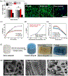
Morphology, composition, and shear-thinning characteristics of the nanocomposite hydrogels. (a) TEM image displaying the nSi dispersion in water (scale bar = 100 nm). (b) Image showing the injection of the blue colored hydrogel through a 22 gauge surgical needle. (c) Viscosity vs shear rate plots illustrate an increase in the viscosity due to the presence of nSi. All the formulations display the typical shear-thinning behavior with a reduction in the viscosity as the shear rate increases. (d) Scanning electron microscopy images of the DNA-based hydrogel showing a highly porous structure. The inclusion of nSi reduces the pore size of the hydrogel network (scale bar =100 μm). (e) Measurement of pore diameters by ImageJ, displaying a significant decrease in pore size of the hydrogel network upon addition of nSi. Results are shown as mean ± standard deviation (n = 50) (*p < 0.05, **p < 0.01, ***p < 0.001). (f) Energy-dispersive X-ray spectra of 0% nSi (i.e., DNA gel without nSi) and (g) 0.5% nSi (i.e., DNA gel with 0.5% nSi) indicate the presence of Si and Mg in the nanocomposite hydrogel. (h) XPS of 0.5% nSi hydrogels confirming the presence of nSi in the nanocomposite hydrogel. The Si 2p peak was visible at around 100 eV.
Figure 3.
Figure 3.
Mechanical and structural characterization of nanocomposite hydrogels as injectable materials. (a) Tan δ (G″/G′) profiles for the nanocomposite hydrogels over a range of frequency from 0.01 to 10 Hz. (b) Frequency sweep experiments performed in the range of 0.01 to 10 Hz indicate an increase in storage modulus as the concentration of nSi was increased. (c) Plot of yield stress as a function of nSi concentration. Results are reported as mean ± standard deviation (n = 3) (***p < 0.001). (d) Recovery data obtained by monitoring the storage modulus of the nanocomposite hydrogels while subjecting them to alternating high (100%) and low (1%) strain conditions. Both 0% nSi (i.e., DNA gel without nSi) and 0.5% nSi (i.e., DNA gel with 0.5% nSi) exhibited more than 95% recovery. (e) Temperature sweeps carried out from 25 to 45 °C. Tan δ values displayed no significant changes in the range of experimental temperatures. (f) Fourier transform infrared spectra of the DNA hydrogel with and without nSi. The highlighted bands (asymmetric stretching and bending of phosphate group) indicate the electrostatic interactions between the nanosilicate edges and DNA.
Figure 4.
Figure 4.
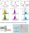
XPS analysis of the nanocomposite DNA-based hydrogel. Comparison of high-resolution XPS spectra of 0% nSi (i.e., DNA gel without nSi) and 0.5% nSi (i.e., DNA gel with 0.5% nSi) for (a) oxygen (O 1s), (b) phosphorus (P 2p), and (c) nitrogen (N 1s). Comparison of the deconvoluted peaks of (d) O 1s, (e) P 2p, and (f) N 1s for both 0% nSi and 0.5% nSi systems. The experimental data points are shown as solid black lines. The rearrangement of the O 1s peak components and the shift of P 2p peak confirmed the presence of attractive electrostatic interactions between the oxygen atom of phosphate anion (PO2) and silicate nanodisks. (g) Proposed mechanism for physical cross-linking between the DNA backbone and silicate nanodisks.
Figure 5.
Figure 5.
In vitro biocompatibility and controlled release properties of the physically cross-linked hydrogels. (a) MTS assay at 24 and 72 h of hASCs in contact with the nanocomposite hydrogels revealed no significant difference in cell viability irrespective of the concentration of nSi used. Cells grown under serum-starved conditions served as a positive control. (b) Representative fluorescence images of actin stained with Alexa Fluor 488 Phalloidin (green) and nuclei stained with DAPI (blue), of hASCs after 72 h of contact with the hydrogels. Cells grown under serum-starved conditions served as a positive control (scale bar = 400 μm). (c) Comparison of release profiles of Dex from DNA hydrogels without nSi and the nanocomposite systems over a period of 10 days. Results are shown as the mean ± standard deviation (n = 3). (d) Half-release time (t1/2) of Dex as a function of nSi concentration confirmed the sustained release behavior of the nanocomposite hydrogels. (e) Viscosity vs shear rate plots of hydrogels loaded with the drug confirming the retention of shear-thinning behavior after Dex loading. (f) Schematic and corresponding images displaying a bone allograft before and after coating with the nanocomposite DNA-based hydrogel. (g) Time-lapse SEM micrographs of the allograft showing the erosion of the coating from the graft surface after 10 days of study (scale bar = 500 μm).
Figure 6.
Figure 6.
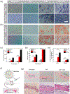
Evaluation of in vitro and in vivo efficacy of the drug-loaded hydrogel for osteogenic differentiation. (a) Images of ALP staining of hASCs after 7 and 14 days of Dex treatment. Both the Dex-treated group (+nSi) and cells grown in osteoinductive media (pos ctrl) show high levels of ALP, representing osteogenic differentiation of hASCs (scale bar = 400 μM). (b) Alizarin red staining of hASCS after 7 and 14 days to detect the calcium deposition (scale bar = 400 μM). Intense red staining signifies osteogenic differentiation of hASCs. (c) ALP quantification demonstrated levels of ALP expression in the Dex-treated group significantly higher than those of the cells grown in basal media (neg ctrl) and osteoconductive media (OC). (d) Quantification of the deposited calcium confirmed significantly higher amounts of calcium in the Dex-treated group as compared to that in neg ctrl and OC. (e) qPCR analysis of different osteogenic markers after 14 days of Dex treatment. The fold expression increase of ALP and COLA1 was assessed following the ΔΔCt relative method. Results are shown as mean ± standard deviation (n = 3) (*p < 0.05, **p < 0.01, ***p < 0.001). Basal media, α-MEM, 10% FBS, 1% penicillin/streptomycin; osteoconductive media, α-MEM, 10% FBS, 1% penicillin/streptomycin, 50 μM ascorbic acid-2-phosphate, 10 mM β-glycerophosphate; osteoinductive media, α-MEM, 10% FBS, 1% penicillin/streptomycin, 50 μM ascorbic acid-2-phosphate, 10 mM β-glycerophosphate, and 10 nM Dex. (f) Schematic representation of the injection of the hydrogel (0.5% nSi + Dex) into a rat cranial defect and a representative image of the hematoxylin and eosin (H&E)-stained rat calvarial defect region. (g) H&E staining of tissue sections obtained from the rat calvarial defects 4 weeks post-treatment. Images were taken at two different magnifications (5× top row and 20× bottom row). Scale bars are 200 μm for the 5× images and 50 μm for the 20× images. The new bone formation in the central region of the cranial defects are marked in the images. The sections enclosed within the dotted lines are magnified in the bottom row images (n = 6).

Similar articles

Cited by

References

    1. Park H; Woo EK; Lee KY Ionically Cross-linkable Hyaluronate-based Hydrogels for Injectable Cell Delivery. J. Controlled Release 2014, 196, 146–153. - PubMed
    1. Tan H; Xiao C; Sun J; Xiong D; Hu X Biological Self-assembly of Injectable Hydrogel as Cell Scaffold via Specific Nucleobase Pairing. Chem. Commun. 2012, 48, 10289–10291. - PubMed
    1. Glassman MJ; Chan J; Olsen BD Reinforcement of Shear Thinning Protein Hydrogels by Responsive Block Copolymer Self-Assembly. Adv. Funct. Mater. 2013, 23, 1182–1193. - PMC - PubMed
    1. Kretlow JD; Young S; Klouda L; Wong M; Mikos AG Injectable Biomaterials for Regenerating Complex Craniofacial Tissues. Adv. Mater. (Weinheim, Ger.) 2009, 21, 3368–3393. - PMC - PubMed
    1. Gaihre B; Uswatta S; Jayasuriya A Reconstruction of Craniomaxillofacial Bone Defects Using Tissue-Engineering Strategies with Injectable and Non-Injectable Scaffolds. J. Funct. Biomater. 2017, 8, 49. - PMC - PubMed

Publication types

MeSH terms

LinkOut - more resources