. 2019 Jan;98(1):117-125.
doi: 10.1177/0022034518796456. Epub 2018 Sep 6.

Serum Amyloid A Contributes to Chronic Apical Periodontitis via TLR2 and TLR4

Affiliations

Serum Amyloid A Contributes to Chronic Apical Periodontitis via TLR2 and TLR4

K Hirai et al. J Dent Res. 2019 Jan.

Abstract

In the current concept of bacterial infections, pathogen-associated molecular patterns (PAMPs) derived from pathogens and damage-associated molecular patterns (DAMPs) released from damaged/necrotic host cells are crucial factors in induction of innate immune responses. However, the implication of DAMPs in apical and marginal periodontitis is unknown. Serum amyloid A (SAA) is a DAMP that is involved in the development of various chronic inflammatory diseases, such as rheumatoid arthritis. In the present study, we tested whether SAA is involved in the pathogenesis of periapical lesions, using human periapical surgical specimens and mice deficient in SAA and Toll-like receptors (TLR). SAA1/2 was locally expressed in human periapical lesions at the mRNA and protein levels. The level of SAA protein appeared to be positively associated with the inflammatory status of the lesions. In the development of mouse periapical inflammation, SAA1.1/2.1 was elevated locally and systemically in wild-type (WT) mice. Although SAA1.1/2.1 double-knockout and SAA3 knockout mice had redundant attenuation of the extent of periapical lesions, these animals showed strikingly improved inflammatory cell infiltration versus WT. Recombinant human SAA1 (rhSAA1) directly induced chemotaxis of WT neutrophils in a dose-dependent manner in vitro. In addition, rhSAA1 stimulation significantly prolonged the survival of WT neutrophils as compared with nonstimulated neutrophils. Furthermore, rhSAA1 activated the NF-κB pathway and subsequent IL-1α production in macrophages in a dose-dependent manner. However, TLR2/TLR4 double deficiency substantially diminished these SAA-mediated proinflammatory responses. Taken together, the SAA-TLR axis plays an important role in the chronicity of periapical inflammation via induction of inflammatory cell infiltration and prolonged cell survival. The interactions of PAMPs and DAMPs require further investigation in dental/oral inflammation.

Keywords: alarmins; chemotaxis; endodontics; inflammation; innate immunity; pattern recognition receptors.


Conflict of interest statement

The authors declare no potential conflicts of interest with respect to the authorship and/or publication of this article.

Figures

Figure 1.
Figure 1.
Serum amyloid A (SAA) was expressed and is present in human periapical lesions. (A) SAA1 and SAA2 gene expressions in 8 human radicular granuloma samples by reverse transcription polymerase chain reaction. (B) Histologic analysis for the localization of SAA1/2. Left panel, representative images of hematoxylin and eosin staining; middle and right panels, immunohistochemistry for SAA (×100). The circle in the middle panel specifies the area for high-power observation (×200) of SAA-positive cells (right panel).
Figure 2.
Figure 2.
Endodontic infection elevated local and systemic levels of SAA in mice as determined by ELISA. (A) Serum levels of SAA1.1/2.1 in WT and SAA 3 KO mice after pulpal infection. *P < 0.05. (B) Local production of SAA in WT periapical lesions. *P < 0.05 vs. day 10. (C) Local production of SAA1.1/2.1 in SAA3 KO lesions. (D) Local production of SAA3 in SAA1.1/2.1 dKO lesions. *P < 0.05 vs. day 10. The level of SAA proteins was expressed as SAA/serum (ng/mL) or SAA/periapical tissue (ng/mg). Error bars indicate SD. dKO, double knockout; KO, knockout; ND, not detected; SAA, serum amyloid A; WT, wild type.
Figure 3.
Figure 3.
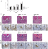
SAA deficiencies attenuate periapical inflammatory cell infiltration but not lesion size. (A) Kinetics of the extent of periapical lesion development in SAA1.1/2.1 dKO, SAA3 KO, and WT mice. The lesion size was obtained by subtraction of an averaged normal periodontal ligament space in baseline controls from the total periapical radiolucent area. Error bar indicates SD. *P < 0.05 vs. WT on day 28. Histology of periapical lesions on (B) day 21 and (C) day 56 after pulp exposure. Original magnification: ×100. In each panel, a representative image of hematoxylin and eosin (HE) staining and immunohistochemistry for neutrophils (Ly-6G) and macrophages (Mac2) is shown. AB, alveolar bone; dKO, double knockout; KO, knockout; R, dental root; SAA, serum amyloid A; WT, wild type.
Figure 4.
Figure 4.
SAA directly induces chemotaxis and survival of neutrophils via TLR2 and TLR4 signaling. (A) Trajectories of WT neutrophils stimulated dose dependently with rhSAA1. (B) Dose-effect of SAA on WT neutrophil chemotaxis. X-axis forward migration index (xFMI) represents the level of the migration directionality. *P < 0.05 vs. nonstimulated cells. (C) Trajectories of TLR-deficient neutrophils stimulated with 100 µg/mL of rhSAA1. (D) Effect of TLR deficiencies on SAA-mediated (100 µg/mL) neutrophil chemotaxis. *P < 0.05 vs. WT. (E) Kinetics of WT neutrophils viability during rhSAA1 stimulation for 18 h. Triplicates per condition. *P < 0.05 vs. control medium. (F) Kinetics of TLR2/4 dKO neutrophils viability during rhSAA1 stimulation for 18 h. Note that in each graph presented in panels A and C, trajectories of 20 randomly selected neutrophils were plotted after correcting the origin. n = 3 per condition in cell viability assays in panels E and F. Error bars in panels B, D, E, and F indicate SD. dKO, double knockout; rhSAA1, recombinant human SAA1; SAA, serum amyloid A; TLR, Toll-like receptor; WT, wild type.
Figure 5.
Figure 5.
rhSAA1 directly stimulates macrophages via TLR2 and TLR4 signaling. (A) SAA3 production by PAMP-stimulated WT macrophages. As macrophages are a major source of extrahepatic SAA3, the effect of PAMPs on SAA3 production was assessed in WT macrophages. SAA3 was induced by activation of LPS-mediated TLR4 signaling but not Pam2CSK4-mediated TLR2 signaling. (B) Dose-effect of rhSAA1 on NF-κB transcriptional activity in NF-κB RAW cells. A mouse macrophage-like cell line, NF-κB RAW cells, was stimulated with rhSAA1 (0.1 to 10 µg/mL), LPS (0.1 µg/mL), Pam2CSK4 (0.1 µg/mL), or control medium. rhSAA1 dose dependently stimulated NF-κB promoter activity, reaching levels comparable to the NF-κB transcriptional activity of LPS (0.1 µg/mL) and Pam2CSK4 (0.1 µg/mL). *P < 0.05 vs. control medium. (C) Effect of TLR2 and TLR4 knockdown on rhSAA1-mediated NF-κB transcriptional activity. Luciferase activity was measured in NF-κB RAW cells treated for 48 h with a scrambled or TLR2- and TLR4-specific siRNA, prior to 6-h treatment with rhSAA1 (1 µg/mL). *P < 0.05 vs. scrambled control siRNA. (D) Dose-effect of rhSAA1 on IL-1α production by WT macrophages. *P < 0.05 vs. control medium. (E) Effect of TLR deficiencies on macrophage IL-1α production stimulated by 100 µg/mL of rhSAA1. n = 3 in all assays. *P < 0.05 vs. WT. Error bars indicate SD. dKO, double knockout; KO, knockout; LPS, lipopolysaccharide; ND, not detected; PAMP, pathogen-associated molecular pattern; rhSAA1, recombinant human SAA1; SAA, serum amyloid A; TLR, Toll-like receptor; WT, wild type.

References

    1. AlShwaimi E, Berggreen E, Furusho H, Rossall JC, Dobeck J, Yoganathan S, Stashenko P, Sasaki H. 2013. IL-17 receptor A signaling is protective in infection-stimulated periapical bone destruction. J Immunol. 191(4):1785–1791. - PMC - PubMed
    1. Anthony D, McQualter JL, Bishara M, Lim EX, Yatmaz S, Seow HJ, Hansen M, Thompson M, Hamilton J, Irving L, et al. 2014. SAA drives proinflammatory heterotypic macrophage differentiation in the lung via CSF-1R-dependent signaling. FASEB J. 28(9):3867–3877. - PMC - PubMed
    1. Ardila CM, Guzmán IC. 2015. Comparison of serum amyloid A protein and C-reactive protein levels as inflammatory markers in periodontitis. J Periodontal Implant Sci. 45(1):14–22. - PMC - PubMed
    1. Bianchi ME. 2007. DAMPs, PAMPs and alarmins: all we need to know about danger. J Leukoc Biol. 81(1):1–5. - PubMed
    1. Bozinovski S, Uddin M, Vlahos R, Thompson M, McQualter JL, Merritt A-S, Wark P, Hutchinson A, Irving L, Levy B, et al. 2012. Serum amyloid A opposes lipoxin A4 to mediate glucocorticoid refractory lung inflammation in chronic obstructive pulmonary disease. Proc Natl Acad Sci U S A. 109(3):935–940. - PMC - PubMed

Publication types

LinkOut - more resources