. 2019 Jan 15:363:11-21.
doi: 10.1016/j.taap.2018.08.024. Epub 2018 Sep 3.

Inhibition of chlorine-induced airway fibrosis by budesonide

Affiliations

Inhibition of chlorine-induced airway fibrosis by budesonide

Sadiatu Musah et al. Toxicol Appl Pharmacol. .

Abstract

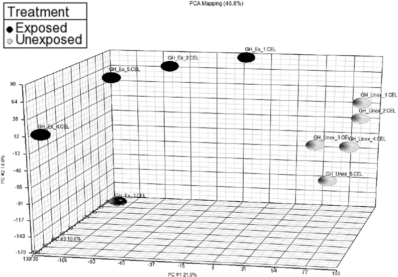
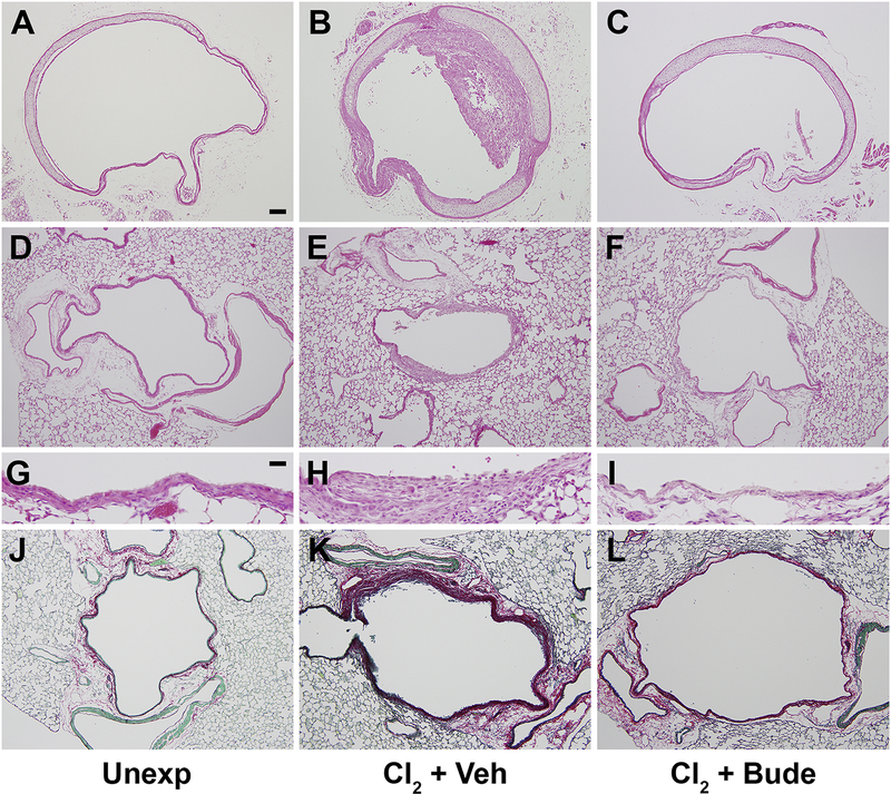
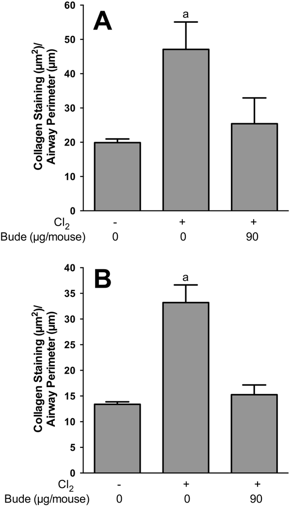
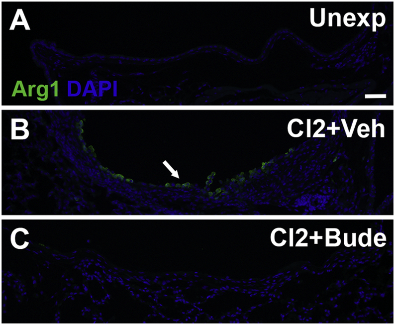
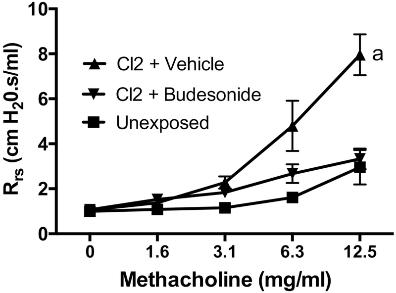
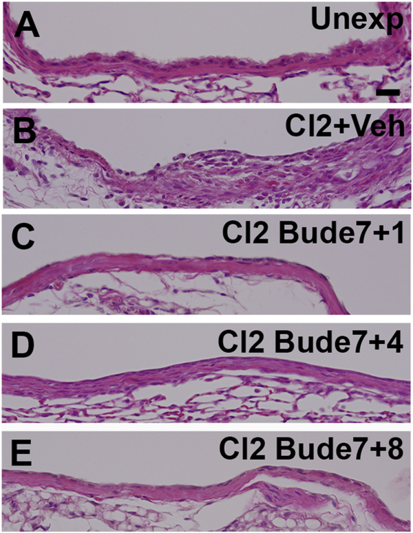
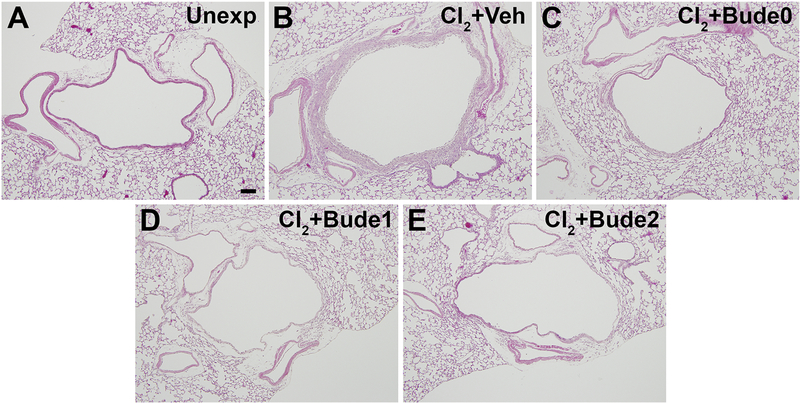
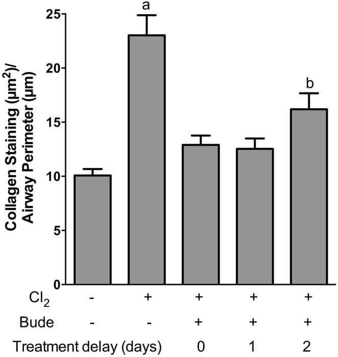
Chlorine is a chemical threat agent that can be harmful to humans. Acute inhalation of high levels of chlorine results in the death of airway epithelial cells and can lead to persistent adverse effects on respiratory health, including airway remodeling and hyperreactivity. We previously developed a mouse chlorine exposure model in which animals developed inflammation and fibrosis in large airways. In the present study, examination by laser capture microdissection of developing fibroproliferative lesions in FVB/NJ mice exposed to 240 ppm-h chlorine revealed upregulation of genes related to macrophage function. Treatment of chlorine-exposed mice with the corticosteroid drug budesonide daily for 7 days (30-90 μg/mouse i.m.) starting 1 h after exposure prevented the influx of M2 macrophages and the development of airway fibrosis and hyperreactivity. In chlorine-exposed, budesonide-treated mice 7 days after exposure, large airways lacking fibrosis contained extensive denuded areas indicative of a poorly repaired epithelium. Damaged or poorly repaired epithelium has been considered a trigger for fibrogenesis, but the results of this study suggest that inflammation is the ultimate driver of fibrosis in our model. Examination at later times following 7-day budesonide treatment showed continued absence of fibrosis after cessation of treatment and regrowth of a poorly differentiated airway epithelium by 14 days after exposure. Delay in the start of budesonide treatment for up to 2 days still resulted in inhibition of airway fibrosis. Our results show the therapeutic potential of budesonide as a countermeasure for inhibiting persistent effects of chlorine inhalation and shed light on mechanisms underlying the initial development of fibrosis following airway injury.

Keywords: Airway fibrosis; Chemical threat agent; Chlorine; Corticosteroid; Inflammation.


Figures

Figure 1.
Figure 1.. Principal component analysis for laser-capture microdissection microarray.
FVB/NJ mice were exposed to chlorine and tracheas were collected 4 days after exposure. Mesenchymal tissue from developing fibrotic lesions was isolated from frozen tracheal sections using laser-capture microdissection. RNA levels were determined by microarray analysis.
Figure 2.
Figure 2.. Effect of chlorine exposure on Arg1 expression.
Chlorine-exposed FVB/NJ mice were euthanized for collection of lungs and trachea 4 or 7 days after exposure. Immunofluorescence staining was performed for Arg 1 in the trachea (A-C) and lobar bronchi (D-F). Arrow shows fibroproliferative lesion with Arg 1 staining in the trachea (in red). Arrowhead shows repaired tracheal epithelium devoid of Arg1 staining. Scale bar in panel C represents 200 μm in A-C and 100 μm in D-F.
Figure 3.
Figure 3.. Effect of budesonide intramuscular treatment on airway fibrosis.
FVB/NJ mice were exposed to chlorine and treated with budesonide or vehicle intramuscularly for 7 days beginning 1 h after exposure. Mice were euthanized for tissue collection 7 days after exposure (1 day after the last budesonide treatment). Hematoxylin and eosin staining was performed in unexposed (A), chlorine-exposed, vehicle-treated (B), and chlorine-exposed, budesonide treated (C) tracheal sections or in lung sections (D-I). PSR staining was performed in lungs from unexposed (J), chlorine-exposed, vehicle treated (K), and chlorine-exposed, budesonide treated (L) mice. Scale bar in panel A represents represent 100 μm in panels A-F and J-L; scale bar in panel G represents 25 μm in panels G-I.
Figure 4.
Figure 4.. Effect of intramuscular budesonide treatment on airway collagen staining.
Chlorine-exposed mice were treated with either vehicle or 90 μg/mouse budesonide for 7 days starting 1 h after exposure. Mice were euthanized for tissue collection 7 days after chlorine exposure. PSR staining was performed on tracheal and lung tissue sections, and quantification of collagen staining was analyzed by ImageJ. (A) Quantification of PSR staining in trachea. a, p<0.05 vs other groups. n=6–8 mice per group. (B) Quantification of PSR staining in lobar bronchi. a, p<0.001 vs other groups. n=6–8 mice per group.
Figure 5.
Figure 5.. Effect of budesonide treatment on Arg1 expression.
Mice were exposed to chlorine and treated with vehicle or budesonide intramuscularly for 7 days starting 1 h after exposure. Mice were euthanized for tissue collection 7 days after exposure. Immunofluorescence staining for Arg 1 staining was performed on lung tissue sections containing lobar bronchi from unexposed (A), chlorine-exposed, vehicle treated (B), and chlorine-exposed, budesonide treated (C) mice. Arrow indicates Arg 1-expressing-cells in airway fibrotic lesion. Scale bar represents 50 μm for all panels.
Figure 6.
Figure 6.. Effect of budesonide treatment on lung function.
Mice were exposed to chlorine and treated with vehicle or budesonide intramuscularly for 7 days starting 1 h after exposure. Lung function was measured 7 days after exposure. a, dose-response curve p<0.05 vs other treatments. n=4–7 mice per group.
Figure 7.
Figure 7.. Airway histology after cessation of budesonide treatment.
Mice were exposed to chlorine and treated with either vehicle or budesonide intramuscularly for 7 days starting 1 h after exposure. Lungs were collected 7, 10, or 14 after exposure (1, 4, or 8 days after cessation of budesonide treatment). Histology of lobar bronchi is shown. Scale bar in panel A represents 25 μm for all panels.
Figure 8.
Figure 8.. Airway collagen staining after cessation of budesonide treatment.
Mice were exposed to chlorine and treated with either vehicle or budesonide intramuscularly for 7 days starting 1 h after exposure. Lungs were collected 7, 10, or 14 after exposure (1, 4, or 8 days after cessation of budesonide treatment). PSR staining was performed on lung tissue sections and collagen staining in lobar bronchi was quantified using ImageJ. a, p<0.01 vs other groups. n=6–8 mice per group.
Figure 9.
Figure 9.. Distribution of airway epithelial cells after cessation of budesonide treatment.
Mice were exposed to chlorine and treated with either vehicle or budesonide intramuscularly for 7 days starting 1 h after exposure. Lungs were collected 7, 10, or 14 after exposure (1, 4, or 8 days after cessation of budesonide treatment). Immunofluorescence staining was performed on lung sections for keratin 5 (basal cell marker), club cell secretory protein (CCSP; club cell marker), and acetylated tubulin (ciliated cell marker). Arrows in panels E and G indicate clusters of K5-stained epithelial cells. Arrow in panel H indicates a cluster of CCSP-stained cells. Arrow in panel J indicates isolated cells that stain for acetylated tubulin. Scale bar in panel A represents 50 μm for all panels.
Figure 10.
Figure 10.. Effect of delayed budesonide treatment on airway fibrosis.
Mice were exposed to chlorine and treated intramuscularly with vehicle or budesonide beginning 1 h after exposure, 1 day after exposure, or 2 days after exposure and treated for 7, 6, or 5 days respectively. Lungs were collected for histological analysis 7 days after chlorine exposure. Hematoxylin and eosin staining of sections containing lobar bronchi are shown. Bude0, Bude1, and Bude2 indicate the start of budesonide treatment either 0 (i.e. the day of exposure), 1, or 2 days after chorine exposure. Scale bar in panel A represents 100 μm for all panels.
Figure 11.
Figure 11.. Effect of delayed budesonide treatment on airway collagen staining.
Mice were exposed to chlorine and treated intramuscularly with vehicle or budesonide beginning 1 h after exposure, 1 day after exposure, or 2 days after exposure and treated for either 7, 6, or 5 days respectively. Lungs were collected for histological analysis 7 days after chlorine exposure. PSR staining was performed on tissue sections, and quantification of collagen staining was performed using ImageJ. a, p<0.01 vs other groups. b, p<0.05 vs unexposed. n=7–9 mice per group.

Similar articles

Cited by

References

    1. Andres SA, Wittliff JL, 2011. Relationships of ESR1 and XBP1 expression in human breast carcinoma and stromal cells isolated by laser capture microdissection compared to intact breast cancer tissue. Endocrine 40, 212–221. - PubMed
    1. Balakrishna S, Song W, Achanta S, Doran SF, Liu B, Kaelberer MM, Yu Z, Sui A, Cheung M, Leishman E, Eidam HS, Ye G, Willette RN, Thorneloe KS, Bradshaw HB, Matalon S, Jordt SE, 2014. TRPV4 inhibition counteracts edema and inflammation and improves pulmonary function and oxygen saturation in chemically induced acute lung injury. Am J Physiol Lung Cell Mol Physiol 307, L158–172. - PMC - PubMed
    1. Bassler D, Plavka R, Shinwell ES, Hallman M, Jarreau PH, Carnielli V, Van den Anker JN, Meisner C, Engel C, Schwab M, Halliday HL, Poets CF, Group NT, 2015. Early Inhaled Budesonide for the Prevention of Bronchopulmonary Dysplasia. N Engl J Med 373, 1497–1506. - PubMed
    1. Bringardner BD, Baran CP, Eubank TD, Marsh CB, 2008. The role of inflammation in the pathogenesis of idiopathic pulmonary fibrosis. Antioxid Redox Signal 10, 287–301. - PMC - PubMed
    1. Chen J, Mo Y, Schlueter CF, Hoyle GW, 2013. Inhibition of chlorine-induced pulmonary inflammation and edema by mometasone and budesonide. Toxicol Appl Pharmacol 272, 408–413. - PMC - PubMed

Publication types

MeSH terms