Review
. 2018 Sep 5;19(9):2628.
doi: 10.3390/ijms19092628.

Actin-Myosin Interaction: Structure, Function and Drug Discovery

Affiliations
Review

Actin-Myosin Interaction: Structure, Function and Drug Discovery

Piyali Guhathakurta et al. Int J Mol Sci. .

Abstract

Actin-myosin interactions play crucial roles in the generation of cellular force and movement. The molecular mechanism involves structural transitions at the interface between actin and myosin's catalytic domain, and within myosin's light chain domain, which contains binding sites for essential (ELC) and regulatory light chains (RLC). High-resolution crystal structures of isolated actin and myosin, along with cryo-electron micrographs of actin-myosin complexes, have been used to construct detailed structural models for actin-myosin interactions. However, these methods are limited by disorder, particularly within the light chain domain, and they do not capture the dynamics within this complex under physiological conditions in solution. Here we highlight the contributions of site-directed fluorescent probes and time-resolved fluorescence resonance energy transfer (TR-FRET) in understanding the structural dynamics of the actin-myosin complex in solution. A donor fluorescent probe on actin and an acceptor fluorescent probe on myosin, together with high performance TR-FRET, directly resolves structural states in the bound actin-myosin complex during its interaction with adenosine triphosphate (ATP). Results from these studies have profound implications for understanding the contractile function of actomyosin and establish the feasibility for the discovery of allosteric modulators of the actin-myosin interaction, with the ultimate goal of developing therapies for muscle disorders.

Keywords: ATP; FRET; actin; drug discovery; fluorescence; heart failure; myosin.


Conflict of interest statement

D.D.T. holds equity in, and serves as President of, Photonic Pharma LLC. This relationship has been reviewed and managed by the University of Minnesota. Photonic Pharma had no role in this study.

Figures
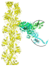
Figure 1
Figure 1
Model, based on superposition of catalytic domain (CD) of myosin crystal structures, for the strong-binding actin-myosin complex (actin yellow, myosin blue, light-chain domain (LCD) down, 2MYS [30]) and weak-binding complex (actin yellow, myosin green, LCD up, 1BR2 [31]). LCD (lever arm) rotation from up to down generates force in the actin-attached power stroke.
Figure 2
Figure 2
Structural states of the skeletal muscle actin-myosin complex detected by TR-FRET. (A) Fluorescence decay of 1 µM AF568 actin (D, black) with 5 µM AF647 labeled S1A1 and 10 µM S1A2 respectively, in the absence (D + A, green) and presence of saturating ATP (D + A + ATP, red), acquired during the steady state. Faster decay in the presence of acceptor indicates FRET. (B) Interprobe distance distribution (best fit to a Gaussian function) corresponding to the bound acto-S1 complex, determined from data in (A) for S (green) and W (red) complexes. (C) Time dependence of FRET-detected mole fractions of the structural states in B, after addition of ATP (at time 0) to a mixture of donor-labeled actin and acceptor-labeled myosin S1. XB (□, black) is the fraction of donor that has bound acceptor. XB = XW + XS, where XW (red) and Xs (green) are the mole fractions of W and S complexes. Adapted from Guhathakurta, P.; Prochniewicz, E.; Thomas, D.D. Amplitude of the actomyosin power stroke depends strongly on the isoform of the myosin essential light chain. Proc. Natl. Acad. Sci. USA 2015, 112, 4660–4665 [26].
Figure 3
Figure 3
Model of actin-myosin complex with skeletal myosin S1 strongly bound to actin. Spheres show the donor (D) and acceptor (A) labeling sites on actin (C374, grey), on myosin (C16, green), and the HCM mutation (E56G, yellow). The C374(actin)-C16(ELC) FRET sensor was designed to determine the effect of the E56G HCM mutation in hVELC on actomyosin function. Adapted from [27], Guhathakurta, P.; Prochniewicz, E.; Roopnarine, O.; Rohde, J.A.; Thomas, D.D. A Cardiomyopathy Mutation in the Myosin Essential Light Chain Alters Actomyosin Structure. Biophys. J. 2017, 113, 91–100, with permission from Elsevier.
Figure 4
Figure 4
Structural states of actomyosin complex affected by a cardiomyopathy mutation as detected by TR-FRET. (A) Time dependence of FRET-detected mole fractions (X) of structural states after addition of ATP to a mixture of donor-labeled actin and acceptor-labeled myosin S1 (WT, top and E56G, bottom). XB (■, black) is the fraction of donor that has bound acceptor. XB = XW + XS, where XW (red) and XS (green) are the mole fractions of W and S complexes. (B) Time dependence of mole fractions (expanded view) in the first minute after addition of ATP, demonstrating a true steady state. Steady-state duty ratio is shown in the box. (C) Interprobe distance (R) and distribution (Γ) (best fit to a Gaussian function) corresponding to bound acto-S1 complex determined from S (green) and W (red) complexes (WT, top and E56G, bottom). While the structure (distance distribution) of the S complex is not significantly affected by the mutation, the W complex is shifted to a shorter distance and a narrower width, so it is designated W′. Each curve is normalized to the unit area, which is independent of the mole fraction XS or XW. Thus the distribution of actin-bound distances R at a given time after mixing is given by a linear combination ρ(R) = (XS/XBS(R) + (XW/XBW(R). Adapted from [27], Guhathakurta, P.; Prochniewicz, E.; Roopnarine, O.; Rohde, J.A.; Thomas, D.D. A Cardiomyopathy Mutation in the Myosin Essential Light Chain Alters Actomyosin Structure. Biophys. J. 2017, 113, 91–100, with permission from Elsevier.
Figure 5
Figure 5
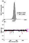
Time-resolved fluorescence resonance energy transfer (TR-FRET) based HTS of National clinical collection (NCC) library for compounds that modulate actin-ANT FRET. (A) Histogram plots of all NCC library compounds after removal of fluorescent compounds show an average FRET efficiency of 0.45 ± 0.003. (B) FRET from a representative NCC screen with “Hit” threshold (>4 SD of mean) indicated by magenta lines. Reproducible “Hits” from triplicate screens are shown in red. Adapted from Guhathakurta, P.; Prochniewicz, E.; Grant, B.D.; Peterson, K.C.; Thomas, D.D. High-throughput screen, using time-resolved FRET, yields actin-binding compounds that modulate actin-myosin structure and function. J. Biol. Chem. 2018, 293, 12288–12298 [28].
Figure 6
Figure 6
High-throughput fluorescence resonance energy transfer (FRET) from actin labeled at the C-terminus (orange spheres) to a peptide derived from the N terminus (green sphere) of the myosin essential light chain, detects actin-binding compounds that change actomyosin structure and function. Adapted from the cover of Guhathakurta, P.; Prochniewicz, E.; Grant, B.D.; Peterson, K.C.; Thomas, D.D. High-throughput screen, using time-resolved FRET, yields actin-binding compounds that modulate actin-myosin structure and function. J. Biol. Chem. 2018, 293, 12288–12298 [28].

Similar articles

Cited by

References

    1. Thomas D.D., Prochniewicz E., Roopnarine O. Changes in actin and myosin structural dynamics due to their weak and strong interactions. Result. Probl. Cell Differ. 2002;36:7–19. - PMC - PubMed
    1. Thomas D.D., Kast D., Korman V.L. Site-directed spectroscopic probes of actomyosin structural dynamics. Annu. Rev. Biophys. 2009;38:347–369. doi: 10.1146/annurev.biophys.35.040405.102118. - DOI - PMC - PubMed
    1. Thomas D.D., Muretta J.M., Colson B.A., Mello R.N., Kast D. Spectroscopic Probes of Muscle Proteins. In: Edward H.E., editor. Comprehensive Biophysics. Elsevier; Amsterdam, The Netherlands: 2012. pp. 226–250.
    1. Sweeney H.L., Houdusse A. Structural and functional insights into the Myosin motor mechanism. Annu. Rev. Biophys. 2010;39:539–557. doi: 10.1146/annurev.biophys.050708.133751. - DOI - PubMed
    1. Wulf S.F., Ropars V., Fujita-Becker S., Oster M., Hofhaus G., Trabuco L.G., Pylypenko O., Sweeney H.L., Houdusse A.M., Schröder R.R. Force-producing ADP state of myosin bound to actin. Proc. Natl. Acad. Sci. USA. 2016;113:E1844–E1852. doi: 10.1073/pnas.1516598113. - DOI - PMC - PubMed

LinkOut - more resources