. 2018 Sep 5;10(9):311.
doi: 10.3390/cancers10090311.

Insonation of Systemically Delivered Cisplatin-Loaded Microbubbles Significantly Attenuates Nephrotoxicity of Chemotherapy in Experimental Models of Head and Neck Cancer

Affiliations

Insonation of Systemically Delivered Cisplatin-Loaded Microbubbles Significantly Attenuates Nephrotoxicity of Chemotherapy in Experimental Models of Head and Neck Cancer

Hang-Kang Chen et al. Cancers (Basel). .

Abstract

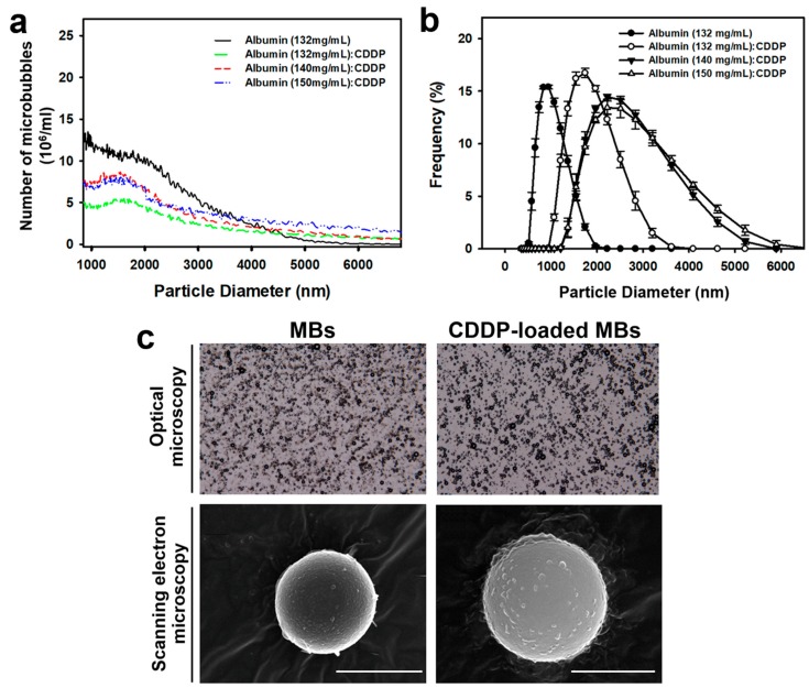
The use of cisplatin (CDDP), the most common chemotherapy drug for head and neck cancer, is limited by its undesirable side effects, especially nephrotoxicity. We investigated ultrasound microbubbles (USMB) as a tool to increase the local intra-tumoral CDDP level while decreasing systemic CDDP cytotoxicity. We allowed CDDP to interact with human serum albumin and then sonicated the resulting CDDP‒albumin complex to generate CDDP-loaded MBs (CDDP-MBs). We then established a head-and-neck tumor-bearing mouse model by implanting FaDu-fLuc/GFP cells into severe combined immunodeficiency mice and used IVIS® bioluminescence imaging to determine the tumor xenograft formation and size. Twice weekly (until Day 33), we administered CDDP only, CDDP + MBs + US, CDDP-MBs, or CDDP-MBs + US intravenously by tail-vein injection. The US treatment was administered at the tumor site immediately after injection. The in vivo systemic distribution of CDDP indicated that the kidney was the most vulnerable organ, followed by the liver, and then the inner ear. However, CDDP uptake into the kidney and liver was significantly decreased in both the CDDP-MBs and CDDP-MBs + US groups, suggesting that MB binding significantly reduced the systemic toxicity of CDDP. The CDDP-MBs + US treatment reduced the tumor size as effectively as conventional CDDP-only chemotherapy. Therefore, the combination of CDDP-MBs with ultrasound is effective and significantly attenuates CDDP-associated nephrotoxicity, indicating a promising clinical potential for this approach.

Keywords: chemotherapy; cisplatin; head and neck cancer; microbubble; nephrotoxicity; ultrasound.


Conflict of interest statement

The authors declare no conflict of interest.

Figures

Figure 1
Figure 1
Quantification and light and scanning electron microscopy images of unloaded MBs (MBs) and cisplatin-loaded MBs (CDDP-MBs). (a) The number-dependent and (b) frequency-dependent size distribution of various MBs. Values are expressed as mean ± SEM (n = 5); (c) Representative light microscopy and scanning electron microscopy images of MBs and CDDP-MBs. Scale bar = 2 μm.
Figure 2
Figure 2
Destruction of ultrasound-targeted MBs and the comparison of in vitro CDDP toxicity on the FaDu cell line in various compositions of CDDP and MBs. (A) MB images and (B) the corresponding image intensities in various groups of acoustic exposure settings; (C) Percentage of cell viability evaluated by WST-1 analysis on FaDu cells 48 h after various ultrasound-targeted MB destruction treatments; (D) Cell viability was assayed by WST-1 after treatment with CDDP, CDDP + MBs, and CDDP-MBs for 48 h; (E) The capacity of the CDDP only, CDDP + MBs, and CDDP-MBs treatments to kill FaDu cells, with or without ultrasound exposure, was evaluated after 48 h of treatment by analyzing the cell viability using the WST-1 assay. Values are expressed as mean ± SEM (n = 5). * p < 0.05, ** p < 0.005.
Figure 3
Figure 3
Influence of MBs and US on the in vivo CDDP accumulation. (A) High frequency US images of albumin MBs in tissue-mimicking agarose phantoms before (control) and after sonication at 1, 2, and 3 W/cm2 for 30 s. The image intensities of MBs are quantified; (B) A series of high-frequency US images that focus on head and neck carcinoma tumor lesions (yellow arrows) showed the presence of MBs (green) within the tumor (white arrows) after their administration via the mouse tail vein, and the disappearance of MBs when after exposure to therapeutic US for 30 s; (C) The CDDP level in vital organs was determined 20 min after a single intravenous administration of chemotherapy using ICP-MS quantitative method. Values are expressed as mean ± SEM (n = 5). * p < 0.05, ** p < 0.005.
Figure 4
Figure 4
In vivo imaging system to monitor tumor growth and chemotherapy outcomes in living mice. (A) FaDu cells stably expressing luciferase and GFP reporter genes (FaDu-fLuc/GFP) were subcutaneously implanted into SCID mice. Four days later, the xenograft tumor-bearing mice were divided to five groups (n = 5 mice per group) including control (saline; Group I) and CDDP-based treatment groups: CDDP only (Group II), CDDP + MBs + US (Group III), CDDP-loaded MBs (CDDP-MBs; Group IV), and CDDP-loaded MBs + US (CDDP-MBs + US; Group V). Mice were repeatedly imaged to record luminescence signals and data were reported as radiance (photons/sec/cm2/steradian) with a color bar. A representative mouse from each of the five groups is shown; (B) Analysis of the IVIS images. Tumor size reductions at the end of treatment (day 33) in each chemotherapy group were 53.02 ± 0.61% (Group II), 62.54 ± 3.83% (Group III), 35 ± 4.46% (Group IV), and 52.29 ± 2.09% (Group V); (C) Effects of 33 days of chemotherapy on body weight gains. Values are expressed as mean ± SEM (n = 5). *** p < 0.0001 (two-way ANOVA with Tukey’s multiple comparison test). NS = not significant; Gp = group.
Figure 5
Figure 5
Effects of various CDDP-based chemotherapies on cell death in target tumor tissues and non-target kidney parenchyma. FaDu xenografted tumors and kidneys of SCID mice were removed for histological examination after various CDDP-based chemotherapies for 33 days. (A) Xenografted tumors were examined by H&E staining and TUNEL assays (original magnification ×400). Marked tumor cell apoptosis was observed in response to the CDDP + MBs + US (Group III) treatment, and to a lesser extent to the CDDP (Group II) and CDDP-MBs + US (Group V) treatments, which both exhibited a similar TUNEL positive staining. The CDDP-MBs group (Group IV) showed less positive staining; (B) CDDP-induced nephrotoxicity, which included congested, shrunken, and degenerated glomeruli (arrows); and (C) necrosis of renal tubular cells and tubular dilatation (arrows) associated with increased leukocyte infiltration (asterisks) were observed following CDDP (Group II) and CDDP + MBs + US (Group III) treatments.
Figure 6
Figure 6
A working model of the gas-filled and CDDP-carrying MBs for the application of USMB in head and neck cancer systemic chemotherapy.
Figure 7
Figure 7
Scheme of animal study design. (A) SCID mice were subcutaneously implanted with FaDu-fLuc/GFP cells (2 × 10⁶ cells/mouse) into the right flank region on day 0; (B) On day 4, tumor xenograft formation and size were determined by IVIS® bioluminescence images (version 4.4, Caliper Life Sciences, Alameda, CA, USA). Medication with CDDP-loaded MBs was given by intravenous injection via the tail vein, followed by ultrasound exposure directly on the tumor site; (C) Flow chart of the study design. CDDP = cisplatin; IV = intravenous; MBs = microbubbles; US = ultrasound.
Figure 8
Figure 8
Schematic illustration of (A) the preparation of CDDP-loaded MBs and (B) optimization of ultrasound parameters in vitro.

Similar articles

Cited by

References

    1. Ferlay J., Shin H.R., Bray F., Forman D., Mathers C., Parkin D.M. Estimates of worldwide burden of cancer in 2008: Globocan 2008. Int. J. Cancer. 2010;127:2893–2917. doi: 10.1002/ijc.25516. - DOI - PubMed
    1. Argiris A., Karamouzis M.V., Raben D., Ferris R.L. Head and neck cancer. Lancet. 2008;371:1695–1709. doi: 10.1016/S0140-6736(08)60728-X. - DOI - PMC - PubMed
    1. Paken J., Govender C.D., Pillay M., Sewram V. Cisplatin-associated ototoxicity: A review for the health professional. J. Toxicol. 2016;2016:1809394. doi: 10.1155/2016/1809394. - DOI - PMC - PubMed
    1. Sasaki N., Ishi K., Kudo N., Nakayama S.M.M., Nakamura K., Morishita K., Ohta H., Ishizuka M., Takiguchi M. Spatial and temporal profile of cisplatin delivery by ultrasound-assisted intravesical chemotherapy in a bladder cancer model. PLoS ONE. 2017;12:e0188093. doi: 10.1371/journal.pone.0188093. - DOI - PMC - PubMed
    1. Lammertink B.H.A., Bos C., van der Wurff-Jacobs K.M., Storm G., Moonen C.T., Deckers R. Increase of intracellular cisplatin levels and radiosensitization by ultrasound in combination with microbubbles. J. Control. Release. 2016;238:157–165. doi: 10.1016/j.jconrel.2016.07.049. - DOI - PubMed