. 2018 Sep 5;23(9):2264.
doi: 10.3390/molecules23092264.

Involvement of Up-Regulation of DR5 Expression and Down-Regulation of c-FLIP in Niclosamide-Mediated TRAIL Sensitization in Human Renal Carcinoma Caki Cells

Affiliations

Involvement of Up-Regulation of DR5 Expression and Down-Regulation of c-FLIP in Niclosamide-Mediated TRAIL Sensitization in Human Renal Carcinoma Caki Cells

Jeong Mi Yun et al. Molecules. .

Abstract

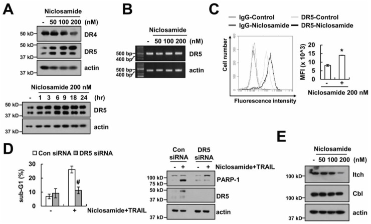
Niclosamide is used to treat intestinal parasite infections, as being an anthelmintic drug. Recently, several papers suggest the niclosamide inhibits multiple signaling pathways, which are highly activated and mutated in cancer. Here, niclosamide was evaluated for identifying strategies to overcome tumor necrosis factor-related apoptosis-inducing ligand (TRAIL) resistance. Although niclosamide (100⁻200 nM) alone did not bring about cell death, combinations of niclosamide and TRAIL led to apoptotic cell death in carcinoma cells, but not in normal cells. Niclosamide markedly increased DR5 protein levels, including cell-surface DR5, and decreased c-FLIP protein levels. Down-regulation of DR5 by specific small interfering RNA (siRNA) and ectopic expression of c-FLIP markedly blocked niclosamide plus TRAIL-induced apoptosis. Our findings provide that niclosamide could overcome resistance to TRAIL through up-regulating DR5 on the cell surface and down-regulating c-FLIP in cancer cells. Taken together, niclosamide may be an attractive candidate to overcome TRAIL resistance.

Keywords: DR5; TRAIL; apoptosis; c-FLIP; niclosamide.


Conflict of interest statement

The authors declare no conflicts of interest.

Figures

Figure 1
Figure 1
Niclosamide combined with TRAIL enhances apoptosis in Caki cells. (A) Apoptosis levels were detected by flow cytometry in Caki cells after treatment with niclosamide (100, 200 nM) and/or TRAIL (30, 50 ng/mL) for 24 h. (BE) Representative image (B), 4′,6′-diamidino-2-phenylindole (DAPI) image (C), DNA fragmentation assay (D), and caspase activity assay (E) were determined by microscopy or assay kit in Caki cells after incubation with 200 nM niclosamide and/or 50 ng/mL TRAIL for 24 h. (F) Apoptosis levels were detected by flow cytometry in Caki cells after pretreatment with 20 μM z-VAD-fmk (z-VAD) for 30 min, followed by treatment with 200 nM niclosamide and 50 ng/mL TRAIL for 24 h. Western blotting detects the protein expression (A,F). The values in graph (A,DF) represent the mean ± SEM of three independent samples. * p < 0.01 compared to the control. # p < 0.01 compared to the niclosamide plus TRAIL.
Figure 2
Figure 2
Niclosamide decreases levels of c-FLIP expression. Protein expression was detected by Western blotting in Caki cells after treatment with niclosamide (50–200 nM) for 24 h.
Figure 3
Figure 3
The down-regulation of c-FLIP is associated with the reduction of TRAIL resistance by niclosamide. (A) The messenger RNA (mRNA) expression was determined by reverse transcription polymerase chain reaction (RT-PCR) (upper panel) and quantitative polymerase chain reaction (qPCR) (lower panel) in Caki cells after treatment with niclosamide (50–200 nM) for 24 h. (B) The protein expression was determined by Western blotting in Caki cells after treatment with 200 nM niclosamide in the presence or absence of 20 μg/mL cycloheximide (CHX) for 3–18 h. The band intensity was measured using ImageJ. (C) Apoptosis levels and protein expression were determined by flow cytometry and Western blotting in Vector cells (Caki/Vec) and in c-FLIP-overexpressed cells (Caki/c-FLIP) after treatment with 200 nM niclosamide, and/or 50 ng/mL TRAIL for 24 h. The values in graph (A,C) represent the mean ± SEM of three independent samples. * p < 0.01 compared to niclosamide plus TRAIL-treated Caki/Vector.
Figure 4
Figure 4
The up-regulation of DR5 by niclosamide is involved in TRAIL sensitization. (A,B) The protein and mRNA expression was determined by Western blotting (A) and RT-PCR (B) in Caki cells after treatment with niclosamide (50–200 nM) for 24 h. (C) The cell surface expression level of DR5 was measured by flow cytometry in Caki cells after treatment with 200 nM niclosamide for 24 h. (D) Apoptosis levels and protein expressions were determined by flow cytometry and Western blotting in Caki cells after transfection with control siRNA (Con siRNA) and DR5 siRNA, followed by treatment with 200 nM niclosamide plus 50 ng/mL TRAIL for 24 h. (E) The protein expression was determined by Western blotting in Caki cells after treatment with niclosamide (50–200 nM) for 24 h. The values in graph (C,D) represent the mean ± SEM of three independent samples. * p < 0.01 compared to the control. # p < 0.01 compared to the niclosamide plus TRAIL-treated control siRNA.
Figure 5
Figure 5
The effects of the niclosamide plus TRAIL on apoptosis in other cancer and normal cells. (A,B) Apoptosis levels and protein expressions were determined by flow cytometry and Western blotting in ACHN, A498, and SK-Hep1 cells after treatment with 200 nM niclosamide and/or 50 ng/mL TRAIL for 24 h. (C) Protein expression was determined by Western blotting in ACHN, A498, and SK-Hep1 cells after treatment with niclosamide (50–200 nM) for 24 h. (D) Representative image and apoptosis levels were detected by interference light microscopy and flow cytometry in Caki, TCMK-1, and HSF cells after treatment with 200 nM niclosamide and/or 50 ng/mL TRAIL for 24 h. The values in the graph (A,D) represent the mean ± SEM of three independent samples. * p < 0.01 compared to the control.
Figure 6
Figure 6
Niclosamide plus TRAIL-induced apoptosis is independent of endoplasmic reticulum (ER) stress and reactive oxygen species (ROS) generation. (A) The protein expression were determined by Western blotting in Caki cells after treatment with niclosamide (50–200 nM) or 2 μM brefeldin A (positive control; p.c.) for 9 h. (B) The apoptosis levels and protein expression were detected by flow cytometry and Western blotting in Caki cells after pretreatment with 5 mM NAC, 200 μM trolox, and 2 mM GEE for 30 min, followed by treatment with 200 nM niclosamide and 50 ng/mL TRAIL for 24 h. The values in graph (B) represent the mean ± SEM of three independent samples.

Similar articles

References

    1. French L.E., Tschopp J. The TRAIL to selective tumor death. Nat. Med. 1999;5:146–147. doi: 10.1038/5505. - DOI - PubMed
    1. Wang S., El-Deiry W.S. TRAIL and apoptosis induction by TNF-family death receptors. Oncogene. 2003;22:8628–8633. doi: 10.1038/sj.onc.1207232. - DOI - PubMed
    1. LeBlanc H.N., Ashkenazi A. Apo2L/TRAIL and its death and decoy receptors. Cell Death Differ. 2003;10:66–75. doi: 10.1038/sj.cdd.4401187. - DOI - PubMed
    1. Ashkenazi A. Targeting death and decoy receptors of the tumour-necrosis factor superfamily. Nat. Rev. Cancer. 2002;2:420–430. doi: 10.1038/nrc821. - DOI - PubMed
    1. Chawla-Sarkar M., Bae S.I., Reu F.J., Jacobs B.S., Lindner D.J., Borden E.C. Downregulation of Bcl-2, FLIP or IAPs (XIAP and survivin) by siRNAs sensitizes resistant melanoma cells to Apo2L/TRAIL-induced apoptosis. Cell Death Differ. 2004;11:915–923. doi: 10.1038/sj.cdd.4401416. - DOI - PubMed

MeSH terms

Substances

LinkOut - more resources