

Improved Chondrogenic Differentiation of rAAV SOX9-Modified Human MSCs Seeded in Fibrin-Polyurethane Scaffolds in a Hydrodynamic Environment
- PMID: 30189664
- PMCID: PMC6163252
- DOI: 10.3390/ijms19092635
Improved Chondrogenic Differentiation of rAAV SOX9-Modified Human MSCs Seeded in Fibrin-Polyurethane Scaffolds in a Hydrodynamic Environment
Abstract
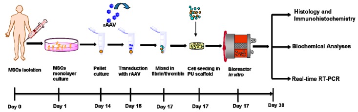
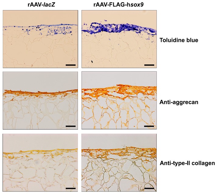
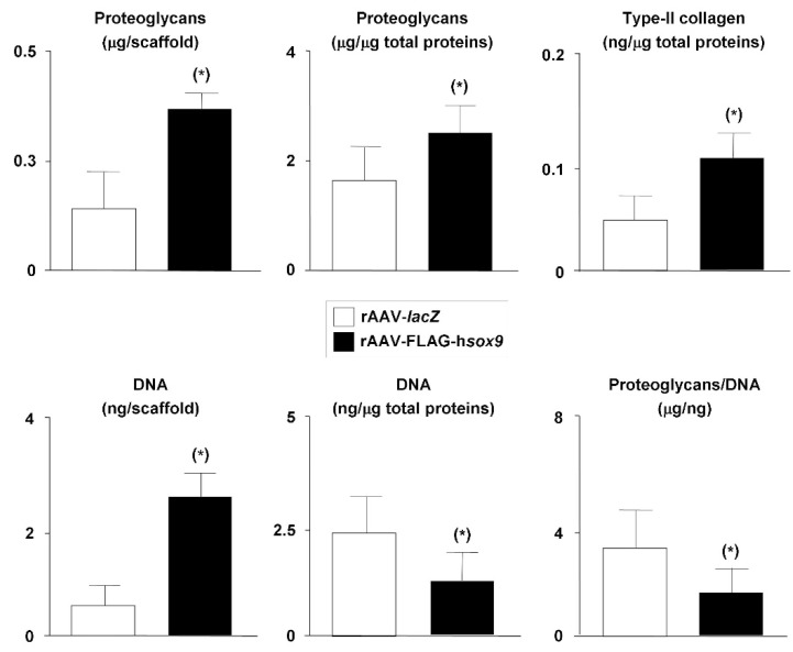
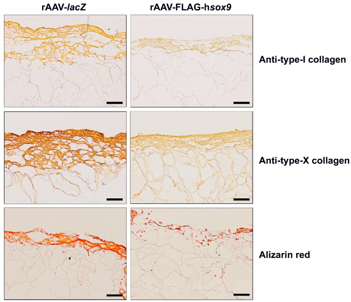
The repair of focal articular cartilage defects remains a problem. Combining gene therapy with tissue engineering approaches using bone marrow-derived mesenchymal stem cells (MSCs) may allow the development of improved options for cartilage repair. Here, we examined whether a three-dimensional fibrin-polyurethane scaffold provides a favorable environment for the effective chondrogenic differentiation of human MSCs (hMSCs) overexpressing the cartilage-specific SOX9 transcription factor via recombinant adeno-associated virus (rAAV) -mediated gene transfer cultured in a hydrodynamic environment in vitro. Sustained SOX9 expression was noted in the constructs for at least 21 days, the longest time point evaluated. Such spatially defined SOX9 overexpression enhanced proliferative, metabolic, and chondrogenic activities compared with control (reporter lacZ gene transfer) treatment. Of further note, administration of the SOX9 vector was also capable of delaying premature hypertrophic and osteogenic differentiation in the constructs. This enhancement of chondrogenesis by spatially defined overexpression of human SOX9 demonstrate the potential benefits of using rAAV-modified hMSCs seeded in fibrin-polyurethane scaffolds as a promising approach for implantation in focal cartilage lesions to improve cartilage repair.
Keywords: SOX9; bioreactors; cartilage repair; chondrogenesis; fibrin-polyurethane scaffolds; hMSCs; rAAV.
Conflict of interest statement
The authors declare that they have no conflicts of interest.
Figures


Similar articles
-
Chondrogenic Differentiation Processes in Human Bone-Marrow Aspirates Seeded in Three-Dimensional-Woven Poly(ɛ-Caprolactone) Scaffolds Enhanced by Recombinant Adeno-Associated Virus-Mediated SOX9 Gene Transfer.Hum Gene Ther. 2018 Nov;29(11):1277-1286. doi: 10.1089/hum.2017.165. Epub 2018 Jun 11. Hum Gene Ther. 2018. PMID: 29717624 Free PMC article.
-
Co-overexpression of TGF-β and SOX9 via rAAV gene transfer modulates the metabolic and chondrogenic activities of human bone marrow-derived mesenchymal stem cells.Stem Cell Res Ther. 2016 Feb 1;7:20. doi: 10.1186/s13287-016-0280-9. Stem Cell Res Ther. 2016. PMID: 26830674 Free PMC article.
-
SOX9 gene transfer via safe, stable, replication-defective recombinant adeno-associated virus vectors as a novel, powerful tool to enhance the chondrogenic potential of human mesenchymal stem cells.Stem Cell Res Ther. 2012;3(3):22. doi: 10.1186/scrt113. Stem Cell Res Ther. 2012. PMID: 22742415 Free PMC article.
-
Regulation and function of SOX9 during cartilage development and regeneration.Semin Cancer Biol. 2020 Dec;67(Pt 1):12-23. doi: 10.1016/j.semcancer.2020.04.008. Epub 2020 May 4. Semin Cancer Biol. 2020. PMID: 32380234 Review.
-
The Role of Physical Stimuli on Calcium Channels in Chondrogenic Differentiation of Mesenchymal Stem Cells.Int J Mol Sci. 2018 Oct 1;19(10):2998. doi: 10.3390/ijms19102998. Int J Mol Sci. 2018. PMID: 30275359 Free PMC article. Review.
Cited by
-
Genetics in Cartilage Lesions: Basic Science and Therapy Approaches.Int J Mol Sci. 2020 Jul 30;21(15):5430. doi: 10.3390/ijms21155430. Int J Mol Sci. 2020. PMID: 32751537 Free PMC article. Review.
-
Combinations of Hydrogels and Mesenchymal Stromal Cells (MSCs) for Cartilage Tissue Engineering-A Review of the Literature.Gels. 2021 Nov 16;7(4):217. doi: 10.3390/gels7040217. Gels. 2021. PMID: 34842678 Free PMC article. Review.
-
Rational design of biodegradable thermoplastic polyurethanes for tissue repair.Bioact Mater. 2021 Dec 31;15:250-271. doi: 10.1016/j.bioactmat.2021.11.029. eCollection 2022 Sep. Bioact Mater. 2021. PMID: 35386346 Free PMC article. Review.
-
Recent advances in polymeric biomaterials-based gene delivery for cartilage repair.Bioact Mater. 2020 Jul 3;5(4):990-1003. doi: 10.1016/j.bioactmat.2020.06.004. eCollection 2020 Dec. Bioact Mater. 2020. PMID: 32671293 Free PMC article.
-
Different types of cartilage neotissue fabricated from collagen hydrogels and mesenchymal stromal cells via SOX9, TGFB1 or BMP2 gene transfer.PLoS One. 2020 Aug 13;15(8):e0237479. doi: 10.1371/journal.pone.0237479. eCollection 2020. PLoS One. 2020. PMID: 32790806 Free PMC article.
References
-
- Hunziker E.B., Lippuner K., Keel M.J., Shintani N. An educational review of cartilage repair: Precepts & practice–myths & misconceptions–progress & prospects. Osteoarthritis Cartilage. 2015;23:334–350. - PubMed
MeSH terms
Substances
LinkOut - more resources
Full Text Sources
Other Literature Sources
Research Materials
