Rheumatoid meningitis presenting with a stroke-like attack treated with recombinant tissue plasminogen activator: a case presentation
- PMID: 30189853
- PMCID: PMC6126002
- DOI: 10.1186/s12883-018-1143-z
Rheumatoid meningitis presenting with a stroke-like attack treated with recombinant tissue plasminogen activator: a case presentation
Abstract
Background: Rheumatoid meningitis presenting with a stroke-like attack (RMSA) is a rare manifestation of rheumatoid arthritis (RA). When the patients arrive within the time-window for recombinant tissue plasminogen activator (rt-PA) infusion therapy, no diagnostic protocol has been established.
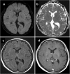
Case presentation: A 55-year-old woman was brought by ambulance to our hospital with complaints of sudden-onset dysarthria and left arm numbness. The National Institutes of Health Stroke Scale (NIHSS) score was 5, and the Alberta Stroke Program Early CT Score was 8. She was diagnosed with acute embolic stroke. At 4 h, 6 min after onset, intravenous administration of rt-PA (alteplase, 0.6 mg/kg) was started. Her neurological deficits improved rapidly, and her NIHSS score was 1. Brain MRI was then performed. There was no hemorrhagic transformation, but the MRI findings were not compatible with ischemic stroke. She had a past history of RA diagnosed 6 months earlier, and she had been treated with methotrexate (10 mg daily). She was diagnosed with RMSA, and continuous infusion of methylprednisolone 1000 mg daily was started for 3 days. The high signal intensity on the FLAIR image disappeared.
Conclusion: CT-based decision-making for rt-PA injection is reasonable, but MRI is needed for the early diagnosis of RMSA. In this case, it is particularly important that neither adverse events nor bleeding complications were observed, suggesting the safety of CT-based thrombolytic therapy in RMSA.
Keywords: Recombinant tissue plasminogen activator (rt-PA); Rheumatoid meningitis; Stroke mimics.
Conflict of interest statement
Ethics approval and consent to participate
Not applicable.
Consent for publication
Written, informed consent was obtained from the patient for the publication of this case report and accompanying images.
Competing interests
The authors declare that they have no competing interests.
Publisher’s Note
Springer Nature remains neutral with regard to jurisdictional claims in published maps and institutional affiliations.
Figures


Similar articles
-
Early neurological deterioration within 24 hours after intravenous rt-PA therapy for stroke patients: the Stroke Acute Management with Urgent Risk Factor Assessment and Improvement rt-PA Registry.Cerebrovasc Dis. 2012;34(2):140-6. doi: 10.1159/000339759. Epub 2012 Aug 1. Cerebrovasc Dis. 2012. PMID: 22854333
-
Tissue plasminogen activator thrombolytic therapy for acute ischemic stroke in 4 hospital groups in Japan.J Stroke Cerebrovasc Dis. 2013 Apr;22(3):190-6. doi: 10.1016/j.jstrokecerebrovasdis.2011.07.016. Epub 2011 Oct 2. J Stroke Cerebrovasc Dis. 2013. PMID: 21968092
-
Intravenous recombinant tissue plasminogen activator therapy for acute ischemic stroke: initial Israeli experience.Isr Med Assoc J. 2004 Feb;6(2):70-4. Isr Med Assoc J. 2004. PMID: 14986460
-
[Intravenous recombinant tissue plasminogen activator in an 18-week pregnant woman with embolic stroke].Rinsho Shinkeigaku. 2010 May;50(5):315-9. doi: 10.5692/clinicalneurol.50.315. Rinsho Shinkeigaku. 2010. PMID: 20535980 Review. Japanese.
-
[Prospects of thrombolytic therapy for acute ischemic stroke].Brain Nerve. 2009 Sep;61(9):1003-12. Brain Nerve. 2009. PMID: 19803399 Review. Japanese.
Cited by
-
Rheumatoid Meningitis Mimicking Clinical and Radiological Findings of Subarachnoid Hemorrhage: A Case Report and Review of the Literature.NMC Case Rep J. 2025 May 20;12:203-208. doi: 10.2176/jns-nmc.2024-0342. eCollection 2025. NMC Case Rep J. 2025. PMID: 40491753 Free PMC article.
-
Rheumatoid meningitis: a rare neurological complication of rheumatoid arthritis.Front Immunol. 2023 Jun 7;14:1065650. doi: 10.3389/fimmu.2023.1065650. eCollection 2023. Front Immunol. 2023. PMID: 37350975 Free PMC article.
-
Rheumatoid arthritis with pachymeningitis - a case presentation and review of the literature.Reumatologia. 2020;58(2):116-122. doi: 10.5114/reum.2020.95368. Epub 2020 Apr 30. Reumatologia. 2020. PMID: 32476685 Free PMC article.
-
Use of Cerebrospinal Fluid Biomarkers in Diagnosis and Monitoring of Rheumatoid Meningitis.Front Neurol. 2019 Jun 26;10:666. doi: 10.3389/fneur.2019.00666. eCollection 2019. Front Neurol. 2019. PMID: 31293505 Free PMC article.
-
Anti-Cyclic Citrullinated Peptide Antibody Index in the Cerebrospinal Fluid for the Diagnosis and Monitoring of Rheumatoid Meningitis.Biomedicines. 2022 Sep 26;10(10):2401. doi: 10.3390/biomedicines10102401. Biomedicines. 2022. PMID: 36289663 Free PMC article.
References
Publication types
MeSH terms
Substances
LinkOut - more resources
Full Text Sources
Other Literature Sources
Medical
Miscellaneous

