Review
. 2018 Nov:71:69-81.
doi: 10.1016/j.dnarep.2018.08.009. Epub 2018 Aug 25.

R-loop generation during transcription: Formation, processing and cellular outcomes

Affiliations
Review

R-loop generation during transcription: Formation, processing and cellular outcomes

Boris P Belotserkovskii et al. DNA Repair (Amst). 2018 Nov.

Abstract

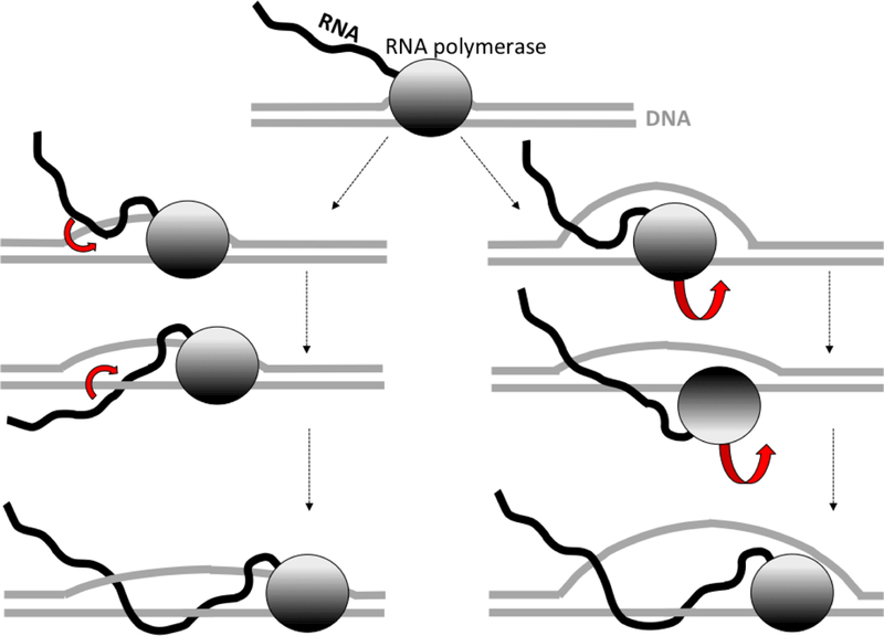
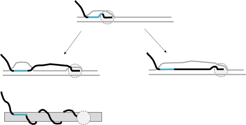
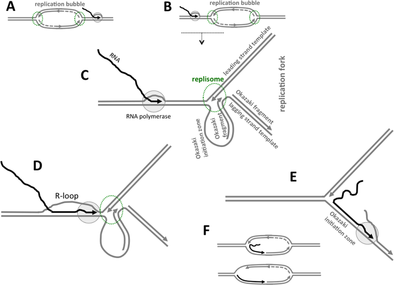
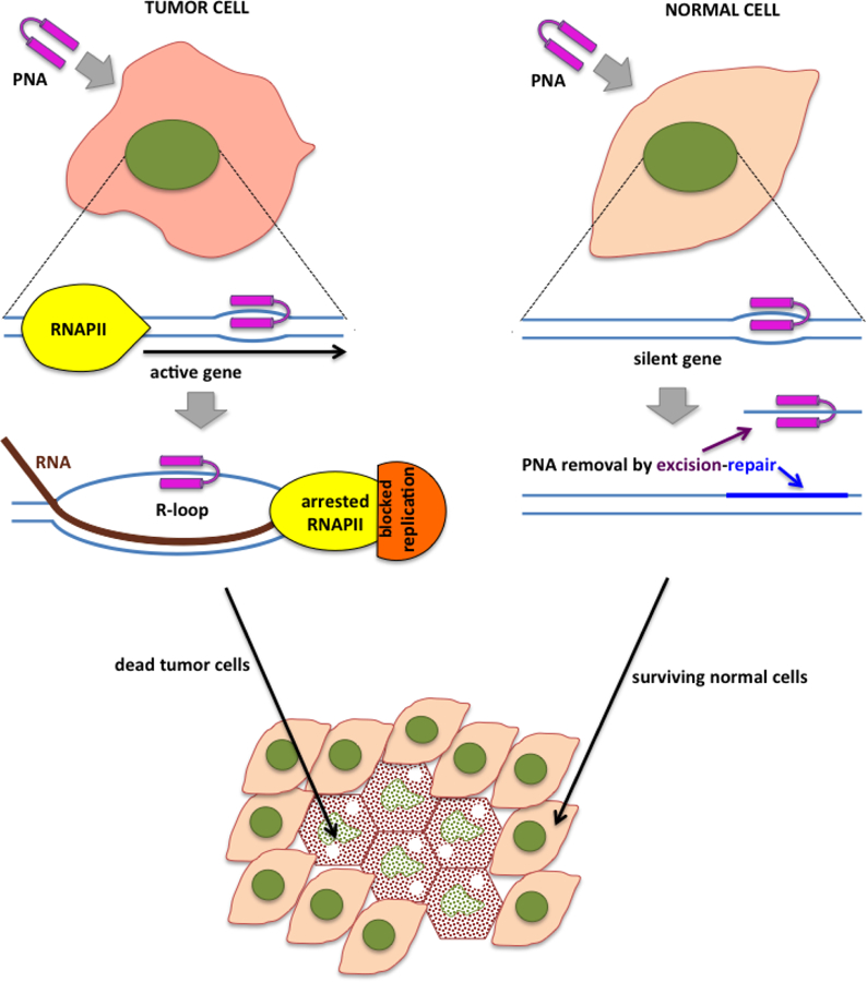
R-loops are structures consisting of an RNA-DNA duplex and an unpaired DNA strand. They can form during transcription upon nascent RNA "threadback" invasion into the DNA duplex to displace the non-template strand. Although R-loops occur naturally in all kingdoms of life and serve regulatory roles, they are often deleterious and can cause genomic instability. Of particular importance are the disastrous consequences when replication forks or transcription complexes collide with R-loops. The appropriate processing of R-loops is essential to avoid a number of human neurodegenerative and other clinical disorders. We provide a perspective on mechanistic aspects of R-loop formation and their resolution learned from studies in model systems. This should contribute to improved understanding of R-loop biological functions and enable their practical applications. We propose the novel employment of artificially-generated stable R-loops to selectively inactivate tumor cells.

Keywords: DNA repair; Non-canonical DNA structures; R-loops; RNA-DNA hybrids; Transcription-replication collisions.


Conflict of interest statement

Conflict of Interest

The authors declare no conflicts of interest

Figures

Fig.1.
Fig.1.. Pathways for R-loop propagation.
DNA is shown in gray, RNA is shown in black, RNAP is shown as a gray oval with color intensity gradient to indicate its orientation. For simplicity, the only inter-strand winding that is shown is the one between the nascent RNA and the template DNA strand. RNA passage and RNAP rotation are indicated by the red block arrows. The pathway at the left shows the intertwining between the nascent RNA and the template DNA strand mediated by the RNA “tail “ passage through the space between the RNA-DNA hybrid and the displaced non-template DNA strand. The pathway at the right shows the intertwining between the nascent RNA and the template DNA strand mediated by RNAP unbinding from the non-template DNA strand followed by rotation around the template DNA strand.
Fig.2.
Fig.2.. Possible modes of behavior for transcription after stable R-loop formation.
DNA is shown in gray lines, RNA is shown in black lines, except the region involved in stable R-loop formation, which is shown in turquoise. RNAP is shown as light gray oval with dashed-line borders. After stable R-loop formation (top), transcriptions could either exit “R-loop mode”(left), or continue in “R-loop mode” (right). Topological scheme below the pathway in the left shows that in this case transcription is associated with the nascent RNA wrapping around DNA.
Fig. 3.
Fig. 3.. R-loop formation upon transcription-replication collisions.
DNA is shown in gray, RNA is shown in black, growing 3’ ends of the nascent DNA or RNA strands are shown by arrows pointed in the direction of growing; RNAP is shown as light-gray oval with black dashed-line borders; replication enzymatic machinery (replisome) is symbolized as an oval area with green dashed-line borders. A: Transcription and replication in co-directional orientation. B: Transcription and replication on the head-on collision course. Replication is usually starts at some small region within the parental DNA duplex called origin, and then the replicated area is expanded forming an unwound region within the parental DNA duplex often referred to as a “replication bubble”. The tip of the replication bubble, which is moving upon the bubble growing, that contains complex multi-protein replication machinery (replisome), is called a “replication fork”. Within the replication fork, one (leading) DNA strand is replicated continuously in the direction of the fork movement, while the other (lagging) DNA strand is replicated discontinuously, forming Okazaki fragments (symbolized by a dashed line). C: More detailed depiction of the region in B underlined by a dashed line with a downward arrow, which shows the replication fork proximal to the transcription complex. The fork is shown in the moment when the synthesis of a new Okazaki fragment (in the loop together with Okazaki initiation zone) is just finished. D: RNAP is stalled upon collision with the replisome that remains intact; and R-loop formation is somehow initiated by RNAP stalling. E: Upon collision with RNAP, the replisome (or its components operating on the lagging DNA strand) dissociate, which allow RNAP to transcribe into single-stranded Okazaki initiation zone, where the likelihood of hybridization between the nascent RNA and the DNA template strand would be very high. Okazaki initiation zone is usually (partially) covered by proteins; however RNAP is probably capable to displace them. F: R-loop-like structures that are formed as a result of pathway in E. Since transcription in R-loop mode is prone to spontaneous blockage, it would probably stop somewhere within the replication bubble. In the bottom structure, the nascent RNA tail invaded into upstream DNA duplex thus extending RNA-DNA hybrid.
Fig. 4.
Fig. 4.. Potential strategy to induce R-loop formation for selective cell suppression.
DNA is shown in blue lines, RNA is shown in brown lines, PNA is shown in magenta.

References

    1. Daube SS and von Hippel PH (1994) RNA displacement pathways during transcription from synthetic RNA-DNA bubble duplexes. Biochemistry, 33, 340347. - PubMed
    1. Yin YW and Steitz TA (2004) The structural mechanism of translocation and helicase activity in T7 RNA polymerase. Cell, 116, 393–404. - PubMed
    1. Jiang M, Ma N, Vassylyev DG and McAllister WT (2004) RNA displacement and resolution of the transcription bubble during transcription by T7 RNA polymerase. Mol Cell, 15, 777–788. - PubMed
    1. Liu X, Bushnell DA and Kornberg RD (2013) RNA polymerase II transcription: structure and mechanism. Biochim Biophys Acta, 1829, 2–8. - PMC - PubMed
    1. Korzheva N and Mustaev A (2001) Transcription elongation complex: structure and function. Curr Opin Microbiol, 4, 119–125. - PubMed

Publication types

LinkOut - more resources