. 2018 Oct 1;215(10):2636-2654.
doi: 10.1084/jem.20171905. Epub 2018 Sep 6.

Remote ischemic post-conditioning promotes hematoma resolution via AMPK-dependent immune regulation

Affiliations

Remote ischemic post-conditioning promotes hematoma resolution via AMPK-dependent immune regulation

Kumar Vaibhav et al. J Exp Med. .

Abstract

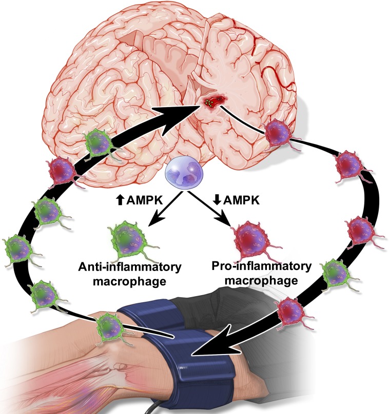
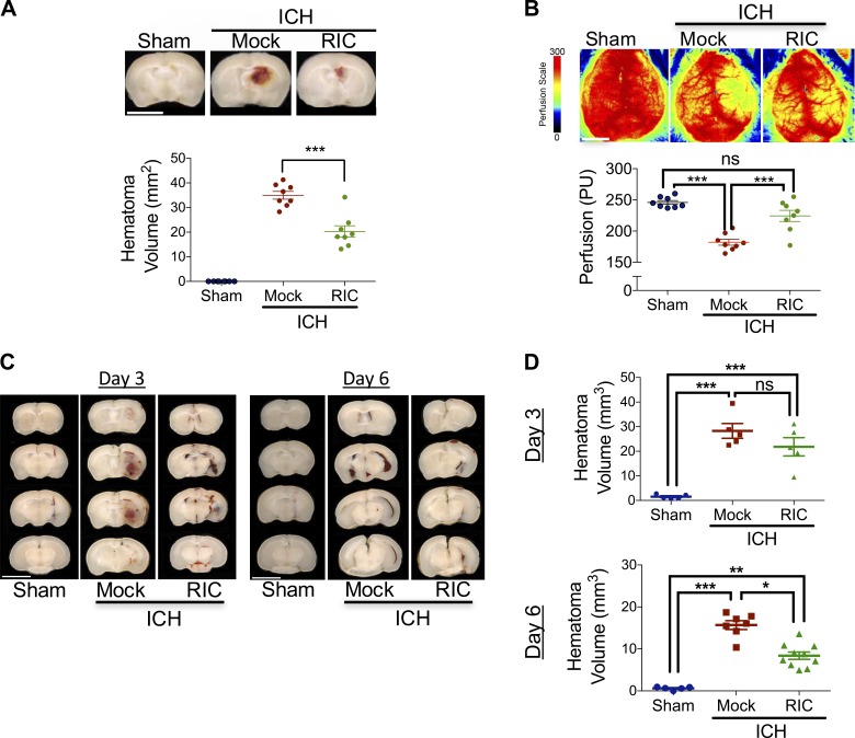
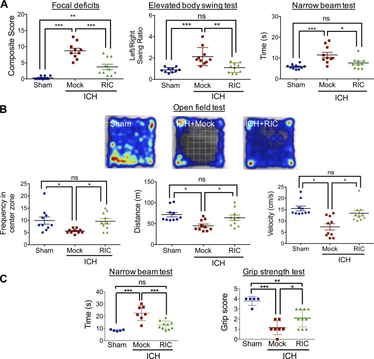
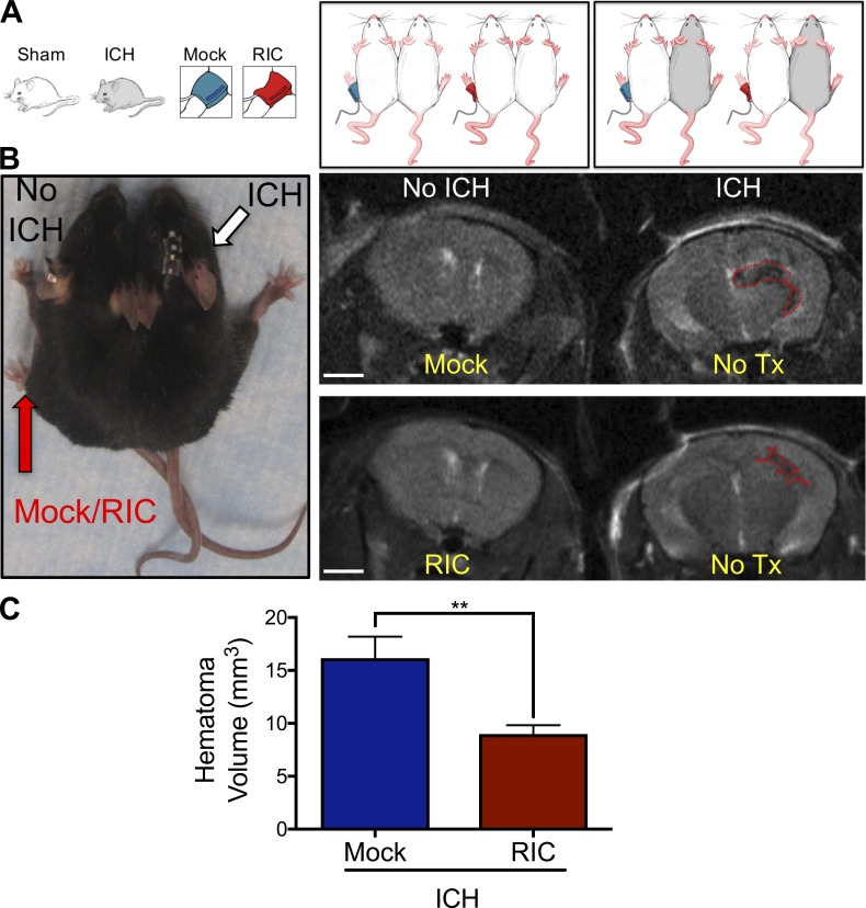
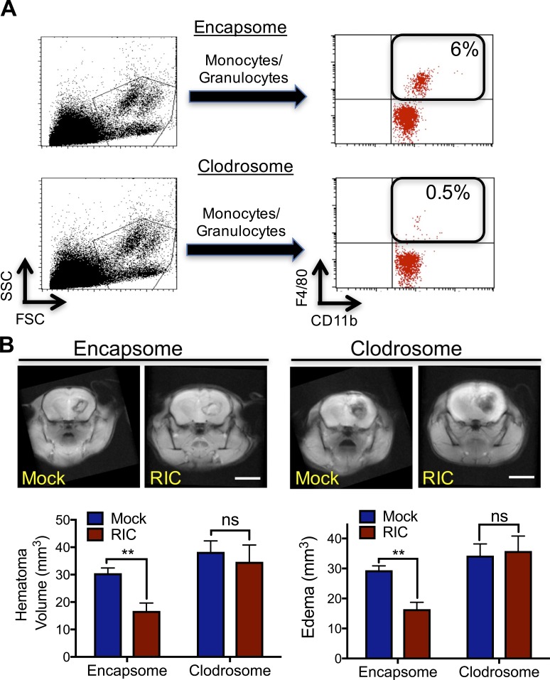
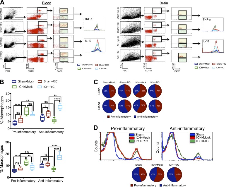
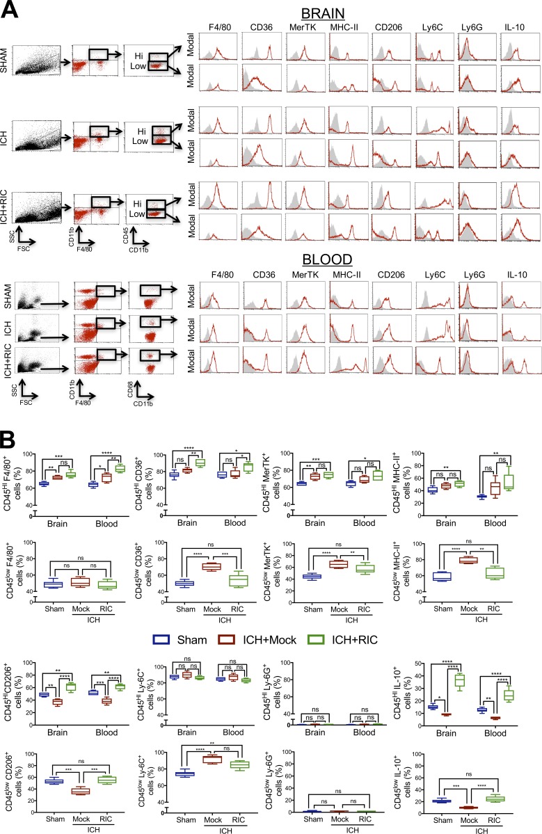
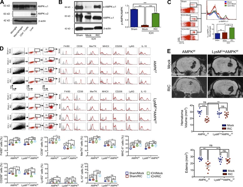
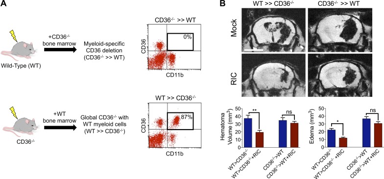
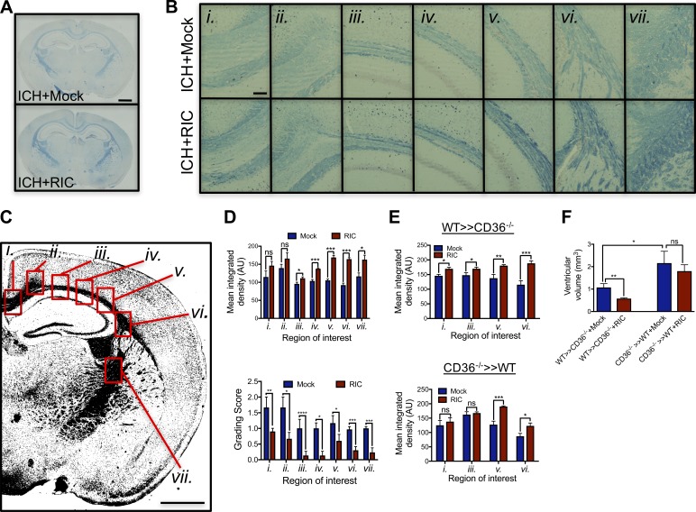
Spontaneous intracerebral hemorrhage (ICH) produces the highest acute mortality and worst outcomes of all stroke subtypes. Hematoma volume is an independent determinant of ICH patient outcomes, making clot resolution a primary goal of clinical management. Herein, remote-limb ischemic post-conditioning (RIC), the repetitive inflation-deflation of a blood pressure cuff on a limb, accelerated hematoma resolution and improved neurological outcomes after ICH in mice. Parabiosis studies revealed RIC accelerated clot resolution via a humoral-mediated mechanism. Whereas RIC increased anti-inflammatory macrophage activation, myeloid cell depletion eliminated the beneficial effects of RIC after ICH. Myeloid-specific inactivation of the metabolic regulator, AMPKα1, attenuated RIC-induced anti-inflammatory macrophage polarization and delayed hematoma resolution, providing a molecular link between RIC and immune activation. Finally, chimera studies implicated myeloid CD36 expression in RIC-mediated neurological recovery after ICH. Thus, RIC, a clinically well-tolerated therapy, noninvasively modulates innate immune responses to improve ICH outcomes. Moreover, immunometabolic changes may provide pharmacodynamic blood biomarkers to clinically monitor the therapeutic efficacy of RIC.


Figures

None
Graphical abstract
Figure 1.
Figure 1.
RIC promotes delayed hematoma resolution and improves outcomes after ICH. (A) Mixed sex C57BL/6J littermates were randomized to receive once-daily mock conditioning or bilateral RIC beginning at 2 h after sham or collagenase-induced ICH. At day 5, hematoma area was quantified in serial 2-mm coronal slices. Bar, 4 mm. Data are mean ± SEM from n = 8 mice/group and were analyzed by one-way ANOVA followed by Tukey’s post-hoc test (***, P < 0.001). Data are representative of two independent experiments. (B) Mixed sex C57BL/6J littermates were randomized to receive once-daily mock conditioning or bilateral RIC beginning at 2 h after sham or collagenase-induced ICH. At day 5, peri-hematoma blood flow was assessed by laser speckle contrast imaging. Bar, 4 mm. Data are mean ± SEM from n = 8 mice/group and were analyzed by one-way ANOVA followed by Tukey’s post-hoc test (***, P < 0.001; ns, not statistically significant). Data are representative of two independent experiments. (C and D) Mixed sex C57BL/6J littermates were randomized to receive once-daily mock conditioning or bilateral RIC beginning at 2 h after sham or intrastriatal placement of 30 µl autologous blood. Littermates were randomized to receive once-daily mock conditioning or bilateral RIC beginning at 2 h after ICH. At day 3 and 6 after injury, hematoma area was quantified in serial 2-mm coronal slices. Bar, 4 mm. Data are mean ± SEM from n = 5–10 mice/group and were analyzed by one-way ANOVA followed by Tukey’s post-hoc test (*, P < 0.05; **, P < 0.01; ***, P < 0.001; ns, not statistically significant). Data are representative of two independent experiments.
Figure 2.
Figure 2.
RIC improves neurobehavioral outcomes after ICH. Mixed sex C57BL/6J littermates were randomized to receive once-daily mock conditioning or bilateral RIC beginning at 2 h after sham or collagenase-induced ICH. (A) Neurological outcomes were assessed at day 4–5 after injury, using a composite focal deficits score (a measure of global injury severity), the elevated body swing test (a task of asymmetric motor behavior), and by the narrow beam test (a measure of motor balance and coordination). Data are mean ± SEM from n = 10 mice/group and were analyzed by one-way ANOVA followed by Tukey’s post-hoc test (*, P < 0.05; **, P < 0.01; ***, P < 0.001; ns, not statistically significant). Data are representative of two independent experiments. (B) Mixed sex C57BL/6J littermates were randomized to receive once-daily mock conditioning or bilateral RIC beginning at 2 h after sham or collagenase-induced ICH. At day 4 after injury, neurological outcomes were assessed using the open field test. Representative heat maps are depicted, and frequency in the center zone, distance traveled, and movement velocity were quantified. Data are mean ± SEM from n = 10 mice/group and were analyzed by one-way ANOVA followed by Tukey’s post-hoc test (*, P < 0.05; ns, not statistically significant). Data are representative of two independent experiments. (C) Mixed sex C57BL/6J littermates were randomized to receive once-daily mock conditioning or bilateral RIC beginning at 2 h after sham or intrastriatal placement of 30 µl autologous blood. Neurobehavioral outcomes, including the narrow beam test and grip strength test, were assessed at day 6 after injury. Data are mean ± SEM from n = 5–10 mice/group and were analyzed by one-way ANOVA followed by Tukey’s post-hoc test (*, P < 0.05; **, P < 0.01; ***, P < 0.001; ns, not statistically significant). Data are representative of two independent experiments.
Figure 3.
Figure 3.
Humoral factors mediate RIC-induced hematoma resolution. (A) Parabiotic pairs (n = 6 pairs/group) were generated using age-matched male C57BL/6J mice and randomized to treatment arms, as illustrated. In all pairs, the left pair mate was treated with either once-daily mock conditioning or RIC for 5 d without injury. The right pair mate was untreated and received either a sham injury or ICH. (B) Photograph of a representative parabiotic pair (left). Mock conditioning or RIC was performed in the left pair mate and sham/ICH was performed on the right pair mate, as indicated. Representative MRI brain scans at day 5 after ICH (right). The brain on the left side is uninjured, and the perimeter of the hematoma is indicated by a red dotted line in the brain on the right side. Bar, 2 mm. (C) Quantification of hematoma volume, as assessed by MRI. Data are mean ± SEM from n = 6 pairs/treatment group. Data are representative of two independent experiments and were analyzed by Student’s t test (**, P < 0.01).
Figure 4.
Figure 4.
Macrophage activation is required for RIC-induced hematoma resolution after ICH. (A) Control liposomes (encapsome) or clodronate liposomes (clodrosome) were administered intraperitoneally to mixed sex C57BL/6J littermates for three consecutive days to deplete myeloid cells. Monocyte/granulocyte populations were identified using FSC/side scatter (SSC) and myeloid cell depletion was quantified in CD11b+, F4/80+ myeloid cells. Inset numbers indicate the percentage of total blood cells. Data are representative of n = 8 mice/group from two independent experiments. (B) Mixed sex C57BL/6J littermates were administered either encapsome or clodrosome for three consecutive days, as indicated in A. Mice were then randomized to receive once-daily mock conditioning or bilateral RIC beginning at 2 h after collagenase-induced ICH. Hematoma volume and edema volume were quantified by MRI at day 5 after ICH. Bar, 4 mm. Data are mean ± SEM from n = 8/group and were analyzed using a Student’s t test (**, P < 0.01; ns, not statistically significant). Data are representative of two independent experiments.
Figure 5.
Figure 5.
RIC promotes anti-inflammatory macrophage polarization. (A and B) CFSE-labeled donor macrophages were intravenously administered (5 × 10⁵ cells/recipient) to male C57BL/6J littermates at 1 h after sham or collagenase-induced ICH. Mice were randomized to receive once-daily mock or RIC beginning at 2 h after injury, and both blood and brain tissue were collected at 72 h after sham/ICH. Live cells were gated according to the FSC/SSC profile and further gating was performed to detect CD11b+, CFSE+ cells populations. The effect of RIC on the phenotype of blood and brain macrophages were further defined as anti-inflammatory (CFSE+, CD11b+, F4/80+, CD206+, IL-10+) or pro-inflammatory (CFSE+, CD11b+, CD206, F4/80+, TNF-α+). Data are mean ± SEM from n = 6 mice/group and were analyzed by one-way ANOVA followed by Tukey’s post-hoc test (**, P < 0.01; ****, P < 0.0001; ns, not statistically significant). Data are representative of two independent experiments. (C) Graphic representation of the ratio of pro-inflammatory/anti-inflammatory macrophages, as determined in B. (D) Representative histograms and graphic representation of the ratio of pro-inflammatory/anti-inflammatory macrophages in blood collected from parabiotic pairs, as defined in A. Data are mean ± SEM from n = 6 pairs/group and are representative of two independent experiments.
Figure 6.
Figure 6.
RIC induces anti-inflammatory macrophage polarization after ICH. (A and B) Mixed sex C57BL/6J littermates were randomized to receive once-daily mock conditioning or bilateral RIC beginning at 2 h after sham or collagenase-induced ICH. Phenotypic analysis of macrophages was performed in peri-hematoma brain tissue (0.2 g) or blood (B; 200 µl) at 72 h after sham, ICH with mock conditioning (ICH), or ICH+RIC. Live cells were gated using FSC/SSC and CD11b+F4/80+ cells were further selected. CD11b+CD45hi-infiltrated myeloid cells and CD11b+CD45low residential myeloid cells were selected from brain tissue, or CD11b+CD68+ macrophages were selected from blood. Selected populations were further defined using F4/80, CD36, MerTK, MHC-II, CD206, Ly-6C, Ly-6G, or IL-10. Representative histograms are provided for each marker. Gray shaded areas indicate isotype controls. Quantified data, which are expressed as the mean ± SEM from n = 6 mice/group, were analyzed using a one-way ANOVA followed by Tukey’s post-hoc test (*, P < 0.05; **, P < 0.01; ***, P < 0.001; ****, P < 0.0001; ns, not statistically significant) and are representative of two independent experiments.
Figure 7.
Figure 7.
Myeloid AMPKα1 mediates hematoma resolution after RIC. (A) Western blot analysis of AMPKα1 and AMPKα2 subunits in microglia, macrophages, CD4+ T cells, and liver (positive control). β-actin was used as a loading control. (B) Phosphorylated AMPKα1/2 (p-AMPKα1/2) and total AMPKα were assessed in brain tissue of male C57BL/6J mice at day 5 after sham/ICH by Western blotting. Densitometry analysis, which are expressed as the mean ± SEM from n = 6 mice/group, were analyzed using a one-way ANOVA followed by Tukey’s post-hoc test (***, P < 0.001; ns, not statistically significant) and are representative of two independent experiments. (C) Phosphorylated AMPKα1 (p-AMPKα1) was assessed in brain tissue of male C57BL/6J mice at day 5 after sham/ICH by flow cytometry. Data are expressed as the mean ± SEM from n = 6 mice/group and were analyzed using a one-way ANOVA followed by Tukey’s post-hoc test (**, P < 0.01; ns, not statistically significant). Data are representative of two independent experiments. (D) Mixed sex WT (AMPKα1f/f) or myeloid-specific AMPKα1 knockout (LysMCreAMPKα1f/f) littermates were randomized to sham/collagenase-induced ICH groups and received either mock conditioning or RIC beginning at 2 h after injury. At 72 h after injury, macrophages were phenotypically defined in peri-hematoma brain tissue (0.2 g) after sham, sham + RIC, ICH with mock conditioning, or ICH+RIC. Live cells were gated using FSC/SSC and CD11b+F4/80+ cells were selected. CD11b+CD45hi-infiltrated macrophages were further defined using F4/80, CD36, MerTK, MHC-II, CD206, Ly-6G, or IL-10. Representative histograms are provided for each marker and isotype controls are indicated by gray shaded areas. Data from two independent experiments are expressed as the mean ± SEM from n = 6 mice/group and were analyzed using a one-way ANOVA followed by Tukey’s post-hoc test (*, P < 0.05; **, P < 0.01; ***, P < 0.001; ****, P < 0.0001; ns, not statistically significant). (E) Mixed sex AMPKα1f/f or LysMCreAMPKα1f/f littermates were randomized to sham/collagenase-induced ICH groups and received either mock conditioning or RIC beginning at 2 h after injury. At day 5 after sham/ICH, hematoma volume and edema were assessed by MRI. Representative images are shown and quantified data are mean ± SEM from n = 8 mice/group. Bar, 4 mm. Data were analyzed using a one-way ANOVA followed by Tukey’s post-hoc test (**, P < 0.01; ***, P < 0.001; ns, not statistically significant) and are representative of three independent experiments.
Figure 8.
Figure 8.
Myeloid CD36 mediates hematoma resolution by RIC. (A) Schematic depicting the generation of myeloid-specific CD36 knockout mice using an irradiation bone marrow chimera approach. Male WT (C57BL/6J) or CD36−/− mice were irradiated and reconstituted using bone marrow from either genotype, as indicated. Flow cytometric analysis of blood (200 µl) collected from mice at 4 wk after bone marrow transplant, showing successful generation of chimeric mice following transplantation of CD36−/− bone marrow into irradiated WT mice (CD36−/− >> WT) or transplantation of WT bone marrow into irradiated CD36−/− mice (WT >> CD36−/−). Inset numbers represent the percentage of CD36+, CD11b+ myeloid cells. (B) WT >> CD36−/− mice and CD36−/− >> WT mice were randomized to sham/ICH groups and received either mock conditioning or RIC. At day 5 after sham/ICH, MRI was used to assess hematoma volume and cerebral edema. Bar, 4 mm. Representative images are depicted and data are expressed as mean ± SEM from n = 8 mice/group. Data were analyzed using a one-way ANOVA followed by Tukey’s post-hoc test (*, P < 0.05; **, P < 0.01; ns = not statistically significant) and are representative of two independent experiments.
Figure 9.
Figure 9.
Myeloid-specific deletion of CD36 increases long-term white matter injury after ICH. (A) Mixed sex C57BL/6J littermates were randomized to receive once-daily mock conditioning or bilateral RIC beginning at 2 h after sham or collagenase-induced ICH. White matter was assessed in coronal brain sections by LFB staining at 8 wk after injury. Bar, 1 mm. (B) Sequential micrographs (20× magnification) demonstrate a sparing effect of RIC on white matter after ICH. Bar, 100 µm. Roman numerals (i.vii.) denote anatomical locations, as indicated in C. Bar, 1 mm. Data are representative of n = 5–7 mice/group from two independent experiments. (D) White matter loss was quantified by mean integrated density and LFB grading score in regions adjacent to the clot location, including the lateral corpus callosum, fimbria, and internal capsule. Representative tissue sections are shown in B (*, P < 0.05; **, P < 0.01; ***, P < 0.001). (E) Male WT >> CD36−/− and CD36−/− >> WT chimera mice were generated, as in Figure 8, randomized to sham/ICH groups, and received either mock conditioning or RIC. At 8 wk after injury, white matter loss was quantified by mean integrated density in selected brain regions. Data are representative of n = 6 mice/group from two independent experiments and were analyzed using a one-way ANOVA followed by Tukey’s post-hoc test (*, P < 0.05; ***, P < 0.001; ns, not statistically significant). (F) Ventricular enlargement, a common manifestation of white matter loss, was quantified by fluid attenuation inversion recovery at 1 mo after ICH in WT >> CD36−/− and CD36−/− >> WT chimera mice. Data are mean ± SEM from n = 5–7/mice group and were analyzed using a one-way ANOVA followed by Tukey’s post-hoc test. Data are representative of two independent experiments.

References

    1. Andreka G., Vertesaljai M., Szantho G., Font G., Piroth Z., Fontos G., Juhasz E.D., Szekely L., Szelid Z., Turner M.S., et al. . 2007. Remote ischaemic postconditioning protects the heart during acute myocardial infarction in pigs. Heart. 93:749–752. 10.1136/hrt.2006.114504 - DOI - PMC - PubMed
    1. Barrett R.J., Hussain R., Coplin W.M., Berry S., Keyl P.M., Hanley D.F., Johnson R.R., and Carhuapoma J.R.. 2005. Frameless stereotactic aspiration and thrombolysis of spontaneous intracerebral hemorrhage. Neurocrit. Care. 3:237–245. 10.1385/NCC:3:3:237 - DOI - PubMed
    1. Bhasin R.R., Xi G., Hua Y., Keep R.F., and Hoff J.T.. 2002. Experimental intracerebral hemorrhage: effect of lysed erythrocytes on brain edema and blood-brain barrier permeability. Acta Neurochir. Suppl. (Wien). 81:249–251. - PubMed
    1. Biswas S.K., and Mantovani A.. 2012. Orchestration of metabolism by macrophages. Cell Metab. 15:432–437. 10.1016/j.cmet.2011.11.013 - DOI - PubMed
    1. Bøtker H.E., Kharbanda R., Schmidt M.R., Bøttcher M., Kaltoft A.K., Terkelsen C.J., Munk K., Andersen N.H., Hansen T.M., Trautner S., et al. . 2010. Remote ischaemic conditioning before hospital admission, as a complement to angioplasty, and effect on myocardial salvage in patients with acute myocardial infarction: a randomised trial. Lancet. 375:727–734. 10.1016/S0140-6736(09)62001-8 - DOI - PubMed

Publication types

Substances