. 2018 Sep 18;115(38):9574-9579.
doi: 10.1073/pnas.1806501115. Epub 2018 Sep 6.

Mechanics of diseased red blood cells in human spleen and consequences for hereditary blood disorders

Affiliations

Mechanics of diseased red blood cells in human spleen and consequences for hereditary blood disorders

He Li et al. Proc Natl Acad Sci U S A. .

Abstract

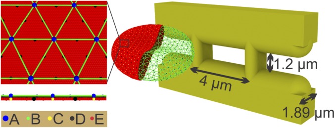
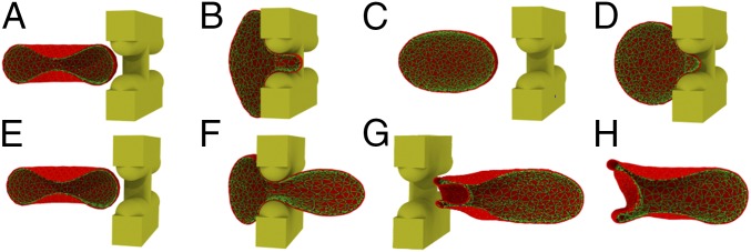
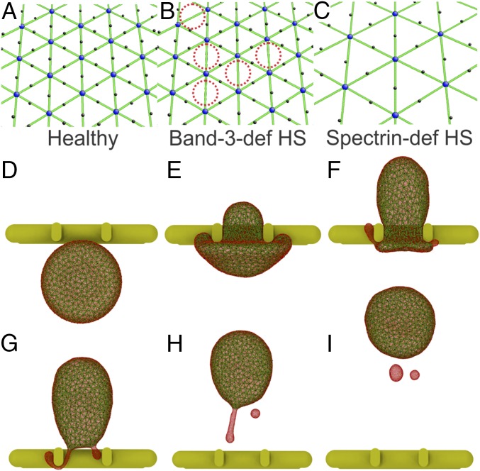
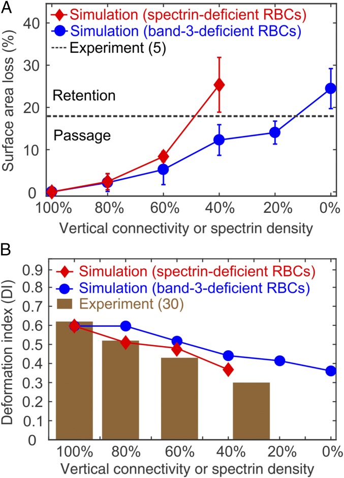
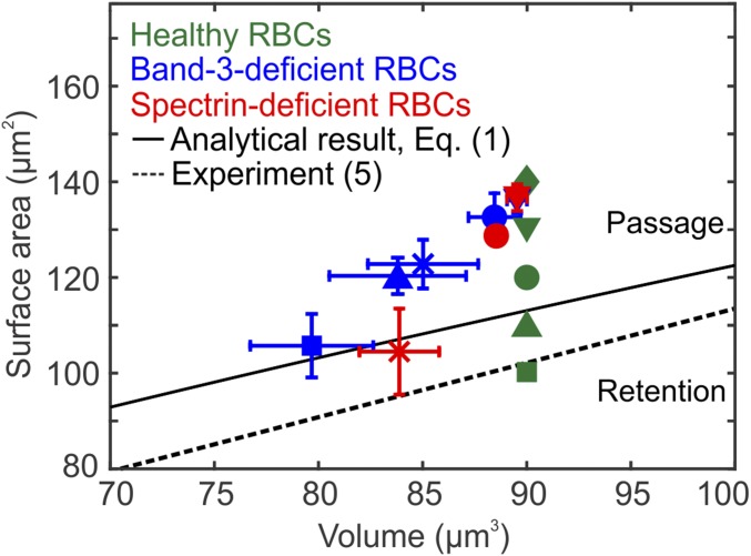
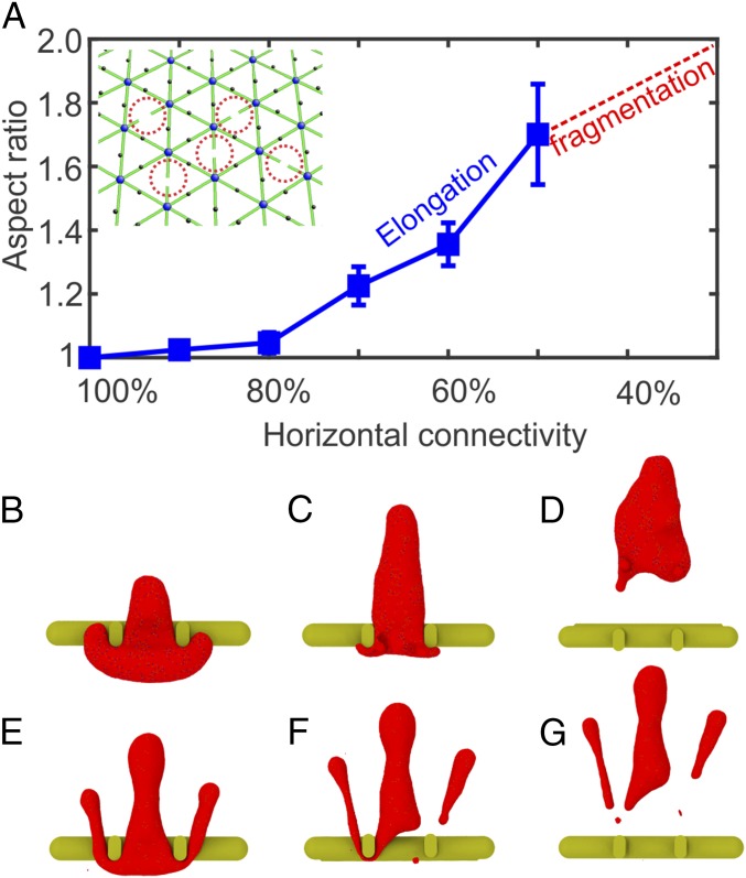
In red blood cell (RBC) diseases, the spleen contributes to anemia by clearing the damaged RBCs, but its unique ability to mechanically challenge RBCs also poses the risk of inducing other pathogenic effects. We have analyzed RBCs in hereditary spherocytosis (HS) and hereditary elliptocytosis (HE), two typical examples of blood disorders that result in membrane protein defects in RBCs. We use a two-component protein-scale RBC model to simulate the traversal of the interendothelial slit (IES) in the human spleen, a stringent biomechanical challenge on healthy and diseased RBCs that cannot be directly observed in vivo. In HS, our results confirm that the RBC loses surface due to weakened cohesion between the lipid bilayer and the cytoskeleton and reveal that surface loss may result from vesiculation of the RBC as it crosses IES. In HE, traversing IES induces sustained elongation of the RBC with impaired elasticity and fragmentation in severe disease. Our simulations thus suggest that in inherited RBC disorders, the spleen not only filters out pathological RBCs but also directly contributes to RBC alterations. These results provide a mechanistic rationale for different clinical outcomes documented following splenectomy in HS patients with spectrin-deficient and ankyrin-deficient RBCs and offer insights into the pathogenic role of human spleen in RBC diseases.

Keywords: cell fragmentation; hereditary elliptocytosis; hereditary spherocytosis; spleen; vesiculation.


Conflict of interest statement

The authors declare no conflict of interest.

Figures

Fig. 1.
Fig. 1.
Simulating RBC passage through IES. The membrane of the RBC is explicitly represented by CG particles. A: actin junctions; B: spectrin particles; C: glycophorin particles; D: band-3 particles; E: lipid particles. The width and height of the simulated slit are 4 μm and 1.2 μm, respectively. The width of the vertical bars is 1 μm, and the thickness of the slit wall is 1.89 μm.
Fig. 2.
Fig. 2.
(A and B) An RBC with surface area of 140 μm2 and volume of 90 μm3 is retained by IES under a pressure gradient of 3 Paμm−1 because of an insufficient driving force. (C and D) An RBC with surface area of 110 μm2 and volume of 90 μm3 is retained by IES under a pressure gradient of 8 Paμm−1 due to reduced surface area. (E–H) Four sequential snapshots of the RBC with surface area of 140 μm2 and volume of 90 μm3 passage through IES under a pressure gradient of 20 Paμm−1 (see Movie S1). Only one half of the RBC is displayed for clarity.
Fig. 3.
Fig. 3.
(A) Cytoskeleton in a healthy RBC model. (B) In the band-3–deficient HS RBC model, the band-3 sites are randomly removed (highlighted by red dotted circles) to represent the effect of band-3 deficiency. (C) In the spectrin-deficient HS RBC model, the density of spectrin network is reduced to represent the effect of spectrin deficiency. (D–I) Six sequential snapshots (top views) of an HS RBC with a vertical connectivity of 60% passage through IES under a pressure gradient of 10 Paμm−1(see Movie S2). Reduced vertical connectivity leads to the detachment of the lipid bilayer from the cytoskeleton and subsequent RBC vesiculation. The lipid CG particles (red particles) are plotted at a smaller size to visualize the cytoskeleton below (green particles).
Fig. 4.
Fig. 4.
(A) Fractional surface area loss of HS RBCs after passage through IES. For the band-3–deficient RBCs, the surface area loss is increased with the decreased vertical connectivity. For the spectrin-deficient RBCs, the surface area loss is increased with the decreased spectrin density. The error bars are computed based on pressure gradient values of 5, 8, 10, 15, and 20 Paμm−1. The black dashed line highlights the critical fraction of surface area loss that determines the retention of RBCs reported by ex vivo experiment (5). (B) DI of the band-3–deficient and the spectrin-deficient RBCs at different levels of HS-related protein deficiency. The brown bar graph shows DI of spectrin-deficient RBCs measured by osmotic gradient ektacytometry at a fixed osmolality of 300 mOsmol/kg (30).
Fig. 5.
Fig. 5.
Prediction of splenic IES retention for healthy RBCs (no protein deficiency) and HS RBCs. Healthy RBCs (green color symbols) with surface area of 140 (), 130(), 120 (), 110 (), and 100 μm2 () and a fixed volume of 90 μm3 are examined, respectively. The surface area and volume of HS RBCs after their passage through IES are plotted. Blue color symbols denote the band-3–deficient RBCs with vertical connectivities of 80% (), 60% (), 40% (×), 20% (), and 0% (). Red color symbols denote the spectrin-deficient RBCs with spectrin density of 80% (), 60% (), and 40% (×). The error bars are computed based on pressure gradient values of 5, 8, 10, 15, and 20 Paμm−1. The black solid and dashed lines highlight the RBC retention threshold predicted by an analytical model given by Eq. 1 (19, 26) and the threshold reported by ex vivo experiment (5), respectively. RBCs with surface area and volume above these thresholds are able to cross IES; otherwise, RBCs are retained by IES.
Fig. 6.
Fig. 6.
(A) Aspect ratios of HE RBCs after passage through IES. When the horizontal connectivity is reduced to 40% or less, HE RBCs break into fragments due to reduced cytoskeleton integrity. The error bars are computed based on pressure gradient values of 5, 8, 10, 15, and 20 Paμm−1. Inset shows that in the spectrin-deficient HE RBC model, spectrin filaments are randomly disassociated (highlighted by red dotted circles), mimicking the disrupted spectrin tetramers. (B–D) Three sequential snapshots (top views) of an HE RBC with a horizontal connectivity of 50% crossing IES (see Movie S3). (E–G) Three sequential snapshots (top views) of an HE RBC with a horizontal connectivity of 20% breaking into fragments after crossing IES (see Movie S4).

References

    1. Mebius RE, Kraal G. Structure and function of the spleen. Nat Rev Immunol. 2005;5:606–616. - PubMed
    1. Petroianu A. The Spleen. Bentham Science Publishers; Hilversum, The Netherlands: 2011.
    1. Buffet PA, et al. Ex vivo perfusion of human spleens maintains clearing and processing functions. Blood. 2006;107:3745–3752. - PubMed
    1. Safeukui I, et al. Retention of plasmodium falciparum ring-infected erythrocytes in the slow, open microcirculation of the human spleen. Blood. 2008;112:2520–2528. - PubMed
    1. Safeukui I, et al. Quantitative assessment of sensing and sequestration of spherocytic erythrocytes by the human spleen. Blood. 2012;120:424–430. - PMC - PubMed

Publication types

MeSH terms

LinkOut - more resources