. 2018 Sep 7;7(9):67.
doi: 10.1038/s41389-018-0078-y.

MEK inhibitors induce apoptosis via FoxO3a-dependent PUMA induction in colorectal cancer cells

Affiliations

MEK inhibitors induce apoptosis via FoxO3a-dependent PUMA induction in colorectal cancer cells

Lin Lin et al. Oncogenesis. .

Retraction in

Abstract

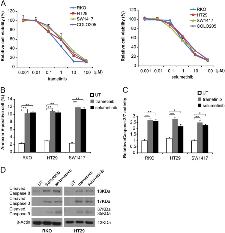
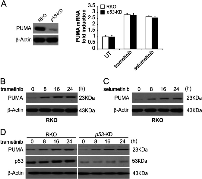
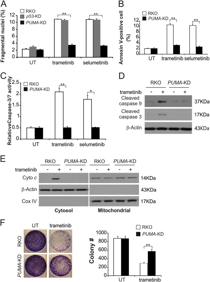
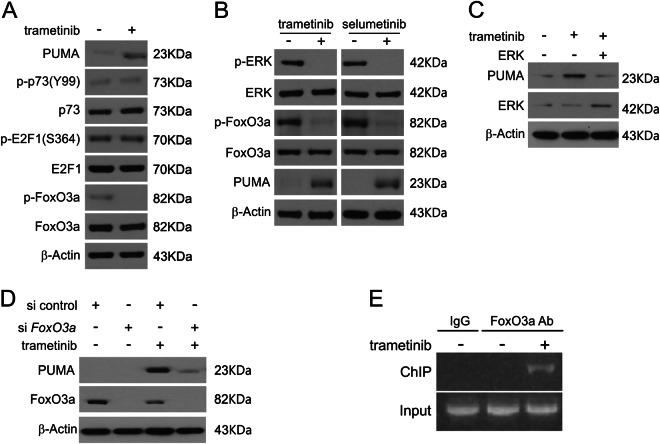
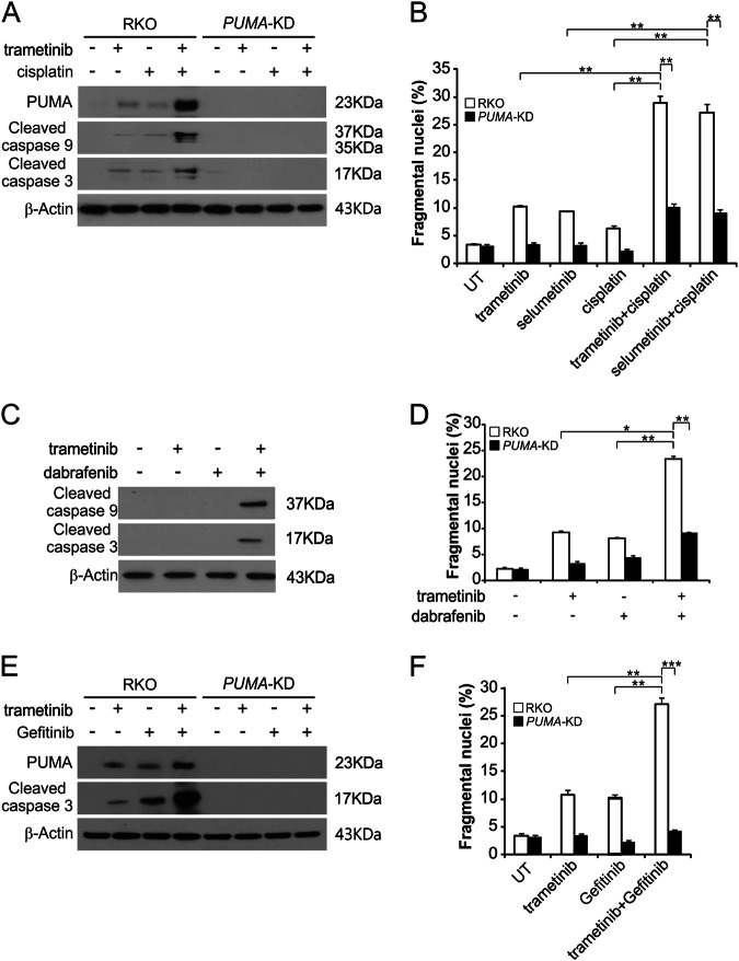
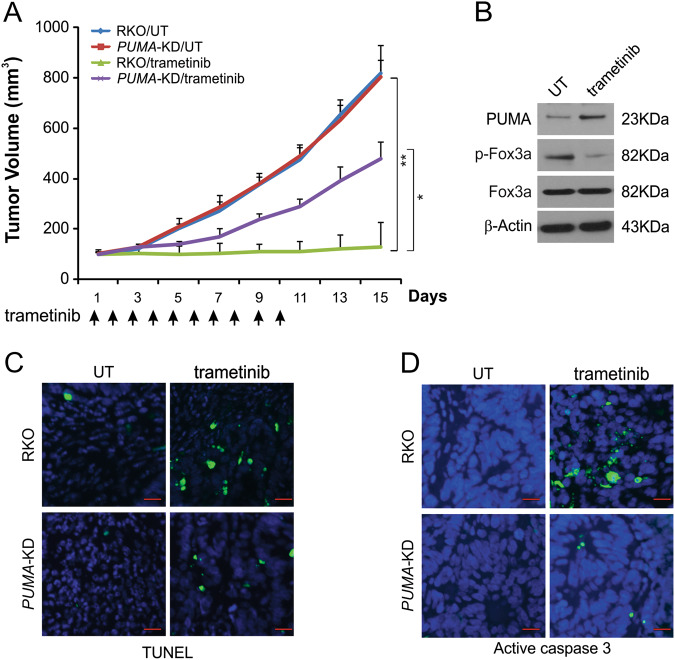
Mutations in BRAF are common to many cancers, including CRC. The MEK inhibitors are being investigated in BRAF-mutant CRC. In this study, we aimed to investigate how MEK inhibitor suppresses growth of BRAF-mutated CRC cells as well as its potential mechanisms. Our findings indicated that MEK inhibitor promote PUMA expression via ERK/FoxO3a signaling pathway. In addition, PUMA induction is essential for MEK inhibitor-induced apoptosis. Moreover, PUMA induction is required for MEK inhibitors to induced apoptosis in combination with cisplatin, dabrafenib, or Gefitinib. Knockdown of PUMA suppressed the anticancer effect of the MEK inhibitor in vivo. Our findings indicate a novel role for PUMA as a regulator of the antitumor effects of MEK inhibitor, suggesting that PUMA induction may modulate MEK inhibitor sensitivity.


Conflict of interest statement

The authors declare that they have no conflict of interest.

Figures

Fig. 1
Fig. 1. MEK inhibitor induces apoptosis in CRC cells.
a The indicated cell lines were treated with increasing doses of trametinib or selumetinib for 72 h. Cell proliferation was determined by MTS assay. b The indicated cell lines were treated with 0.1 μmol/L trametinib or 0.1 μmol/L selumetinib for 24 h at the indicated concentrations. Apoptosis was analyzed by Annexin V/PI staining followed by flow cytometry. c The indicated cell lines were treated with 0.1 μmol/L trametinib or 0.1 μmol/L selumetinib for 24 h at the indicated concentrations. Caspase 3/7 activity was determined by fluorogenic analysis. d The indicated cell lines were treated with 0.1 μmol/L trametinib or 0.1 μmol/L selumetinib at the indicated time points. Cleaved caspase 3, 8, and 9 were analyzed by Western blotting. The results in a, b, and c are expressed as the means ± SD of three independent experiments. **P < 0.01, *P < 0.05
Fig. 2
Fig. 2. MEK inhibitors induce p53-independent PUMA induction.
a Parental and p53-KD RKO cells were treated with 0.1 μmol/L trametinib or 0.1 μmol/L selumetinib for 24 h. PUMA mRNA induction by trametinib was analyzed by real-time reverse transcriptase (RT) PCR, with β-actin as the control. b RKO cells were treated with 0.1 μmol/L trametinib at the indicated time points. PUMA expression was analyzed by Western blotting. c RKO cells were treated with 0.1 μmol/L selumetinib at the indicated time points. PUMA expression was analyzed by Western blotting. d Parental and p53-KD RKO cells were treated with 1 μmol/L trametinib at the indicated time points. PUMA expression was analyzed by Western blotting
Fig. 3
Fig. 3. PUMA is required for the apoptotic activity of MEK inhibitors.
a Parental, p53-KD and PUMA-KD RKO cells were treated with 0.1 μmol/L trametinib or 0.1 μmol/L selumetinib for 24 h. Apoptosis was analyzed by a nuclear fragmentation assay. b Parental and PUMA-KD RKO cells were treated with 0.1 μmol/L trametinib or 0.1 μmol/L selumetinib for 24 h. Apoptosis was analyzed by Annexin V/PI staining followed by flow cytometry. c Parental and PUMA-KD RKO cells were treated with 0.1 μmol/L trametinib or 0.1 μmol/L selumetinib for 24 h. Caspase 3/7 activity was determined by fluorogenic analysis. d Parental and PUMA-KD RKO cells were treated with 0.1 μmol/L trametinib for 24 h. Cleaved caspase 3 and 9 were analyzed by Western blotting. e The cytoplasm and mitochondria were fractionated from parental and PUMA-KD RKO cells treated with 0.1 μmol/L trametinib for 24 h. The distribution of cytochrome c was analyzed by Western blotting. β-Actin and cytochrome oxidase subunit IV (Cox IV) were analyzed as the control for loading and fractionation. f Parental and PUMA-KD RKO cells were treated with 0.1 μmol/L trametinib for 24 h. Colony formation assay was performed by seeding an equal number of treated cells in 12-well plates and then staining attached cells with crystal violet 10 days later. Left, representative pictures of colonies; Right, quantification of colony numbers. The results in a, b, c and f are expressed as the means ± SD of three independent experiments. **P < 0.01, *P < 0.05
Fig. 4
Fig. 4. Induction of PUMA by MEK inhibitors is mediated through ERK inhibition.
a RKO cells were treated with 0.1 μmol/L trametinib for 24 h. PUMA, p73, E2F1, p-FoxO3a and FoxO3a expression were analyzed by Western blotting. b RKO cells were treated with 0.1 μmol/L trametinib or 0.1 μmol/L selumetinib for 24 h. Indicated protein levels were analyzed by Western blotting. c RKO cells were transfected with ERK plasmid for 6 h and then treated with 1 μmol/L trametinib for 24 h. PUMA and ERK expression was analyzed by Western blotting. d RKO cells were transfected with either a control scrambled siRNA or FoxO3a siRNA for 24 h and then treated with 1 μmol/L trametinib for 24 h. FoxO3a and PUMA expression was analyzed by Western blotting. e Chromatin immunoprecipitation (ChIP) was performed using an anti-FoxO3a antibody on RKO cells following trametinib treatment for 12 h. ChIP with the control IgG was used as a control. PCR was carried out using primers surrounding the FoxO3a binding sites in the PUMA promoter
Fig. 5
Fig. 5. MEK inhibitors synergize with cisplatin, dabrafenib or Gefitinib to induce apoptosis via PUMA in CRC cells.
a Parental and PUMA-KD RKO cells were treated with 0.5 μmol/L trametinib, 0.5 μmol/L selumetinib, 20 mg/L cisplatin, or their combination for 24 h. PUMA, cleaved caspase 3 and 9 were analyzed by Western blotting. b Parental and PUMA-KD RKO cells were treated with 0.5 μmol/L trametinib, 0.5 μmol/L selumetinib, 20 mg/L cisplatin, or their combination for 24 h. Apoptosis was analyzed by a nuclear fragmentation assay. c RKO cells were treated with 0.5 μmol/L trametinib, 1 μmol/L dabrafenib, or their combination for 24 h. Cleaved caspase 3 and 9 were analyzed by Western blotting. d Parental and PUMA-KD RKO cells were treated with 0.5 μmol/L trametinib, 1 μmol/L dabrafenib, or their combination for 24 h. Apoptosis was analyzed by a nuclear fragmentation assay. e Parental and PUMA-KD RKO cells were treated with 0.1 μmol/L trametinib, 0.2 μmol/L gefitinib, or their combination for 24 h. PUMA and cleaved caspase 3 were analyzed by Western blotting. f Parental and PUMA-KD RKO cells were treated with 0.1 μmol/L trametinib, 0.2 μmol/L gefitinib, or their combination for 24 h. Apoptosis was analyzed by a nuclear fragmentation assay. The results in b, d and f are expressed as the means ± SD of three independent experiments. ***P < 0.001, **P < 0.01, *P < 0.05
Fig. 6
Fig. 6. PUMA mediates the anticancer effects of trametinib in a xenograft model.
a Nude mice were injected s.c. with 4 × 106 parental and PUMA-KD RKO cells. After 1 week, mice were treated with 1 mg/kg trametinib or buffer for ten consecutive days. The tumor volume at the indicated time points after treatment was calculated and plotted (n = 6 in each group). Arrows indicate trametinib injection. The results are expressed as the means ± SD of three independent experiments. **P < 0.01; *P < 0.05. b Parental RKO xenograft tumors were treated with 1 mg/kg trametinib or the control buffer as in a for four consecutive days. The indicated protein in representative tumors was analyzed by Western blotting. c Paraffin-embedded sections of tumor tissues from mice treated as in b were analyzed by TUNEL staining. d Tissue sections from c were analyzed by active caspase 3 staining. Scale bars 25 μm

References

    1. Solit, D. B. et al. BRAF mutation predicts sensitivity to MEK inhibition. Nature439, 358–362 (2006). - PMC - PubMed
    1. Zhao, Y. & Adjei, A. A. The clinical development of MEK inhibitors. Nat. Rev. Clin. Oncol.11, 385–400 (2014). - PubMed
    1. Luke, J. J., Ott, P. A. & Shapiro, G. I. The biology and clinical development of MEK inhibitors for cancer. Drugs74, 2111–2128 (2014). - PubMed
    1. Holderfield, M., Deuker, M. M., McCormick, F. & McMahon, M. Targeting RAF kinases for cancer therapy: BRAF-mutated melanoma and beyond. Nat. Rev. Cancer14, 455–467 (2014). - PMC - PubMed
    1. Millington, G. W. Mutations of the BRAF gene in human cancer, by Davies et al. (Nature 2002; 417: 949–54). Clin. Exp. Dermatol.38, 222–223 (2013). - PubMed

Publication types