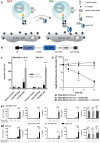
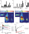
Development of a novel target module redirecting UniCAR T cells to Sialyl Tn-expressing tumor cells
- PMID: 30190468
- PMCID: PMC6127150
- DOI: 10.1038/s41408-018-0113-4
Development of a novel target module redirecting UniCAR T cells to Sialyl Tn-expressing tumor cells
Conflict of interest statement
L.R.L., C.N., M.B., and P.V. have filed patents related to anti-STn mAb L2A5. M.B. has filed patents related to the UniCAR system. M.B. is shareholder of the company GEMoaB which owns the IP related to the UniCAR system. The remaining authors declare that they have no conflict of interest.
Figures


Similar articles
-
TanCAR T cells targeting CD19 and CD133 efficiently eliminate MLL leukemic cells.Leukemia. 2018 Sep;32(9):2012-2016. doi: 10.1038/s41375-018-0212-z. Epub 2018 Jul 25. Leukemia. 2018. PMID: 30046161 No abstract available.
-
Cholesterol Esterification Enzyme Inhibition Enhances Antitumor Effects of Human Chimeric Antigen Receptors Modified T Cells.J Immunother. 2018 Feb/Mar;41(2):45-52. doi: 10.1097/CJI.0000000000000207. J Immunother. 2018. PMID: 29252915
-
Prospects for chimeric antigen receptor-modified T cell therapy for solid tumors.Mol Cancer. 2018 Jan 12;17(1):7. doi: 10.1186/s12943-018-0759-3. Mol Cancer. 2018. PMID: 29329591 Free PMC article. Review.
-
Advances in off-the-shelf CAR T-cell therapy.Clin Adv Hematol Oncol. 2019 Mar;17(3):155-157. Clin Adv Hematol Oncol. 2019. PMID: 30969953 No abstract available.
-
Recent advances in tumor associated carbohydrate antigen based chimeric antigen receptor T cells and bispecific antibodies for anti-cancer immunotherapy.Semin Immunol. 2020 Feb;47:101390. doi: 10.1016/j.smim.2020.101390. Epub 2020 Jan 22. Semin Immunol. 2020. PMID: 31982247 Free PMC article. Review.
Cited by
-
Glycoproteogenomics: Setting the Course for Next-generation Cancer Neoantigen Discovery for Cancer Vaccines.Genomics Proteomics Bioinformatics. 2021 Feb;19(1):25-43. doi: 10.1016/j.gpb.2021.03.005. Epub 2021 Jun 9. Genomics Proteomics Bioinformatics. 2021. PMID: 34118464 Free PMC article. Review.
-
Influence of Culture Conditions on Ex Vivo Expansion of T Lymphocytes and Their Function for Therapy: Current Insights and Open Questions.Front Bioeng Biotechnol. 2022 Jun 29;10:886637. doi: 10.3389/fbioe.2022.886637. eCollection 2022. Front Bioeng Biotechnol. 2022. PMID: 35845425 Free PMC article. Review.
-
UniCAR T cell immunotherapy enables efficient elimination of radioresistant cancer cells.Oncoimmunology. 2020 Apr 5;9(1):1743036. doi: 10.1080/2162402X.2020.1743036. eCollection 2020. Oncoimmunology. 2020. PMID: 32426176 Free PMC article.
-
Extended half-life target module for sustainable UniCAR T-cell treatment of STn-expressing cancers.J Exp Clin Cancer Res. 2020 May 5;39(1):77. doi: 10.1186/s13046-020-01572-4. J Exp Clin Cancer Res. 2020. PMID: 32370811 Free PMC article.
-
CRASH-IT Switch Enables Reversible and Dose-Dependent Control of TCR and CAR T-cell Function.Cancer Immunol Res. 2021 Sep;9(9):999-1007. doi: 10.1158/2326-6066.CIR-21-0095. Epub 2021 Jun 30. Cancer Immunol Res. 2021. PMID: 34193461 Free PMC article.
References
-
- Sadelain M, Brentjens R, Rivière I. The basic principles of chimeric antigen receptor design. Cancer Discov. 2013;3:388–98. doi: 10.1158/2159-8290.CD-12-0548. - DOI - PMC - PubMed
-
- Kochenderfer JN, Dudley ME, Feldman SA, Wilson WH, Spaner DE, Maric I, et al. B-cell depletion and remissions of malignancy along with cytokine-associated toxicity in a clinical trial of anti-CD19 chimeric-antigen-receptor-transduced T cells. Blood. 2012;119:2709–2720. doi: 10.1182/blood-2011-10-384388. - DOI - PMC - PubMed
Publication types
MeSH terms
Substances
LinkOut - more resources
Full Text Sources
Other Literature Sources

