Review
. 2018 Sep 6;4(1):21.
doi: 10.1038/s41572-018-0018-3.

Wilson disease

Affiliations
Review

Wilson disease

Anna Członkowska et al. Nat Rev Dis Primers. .

Abstract

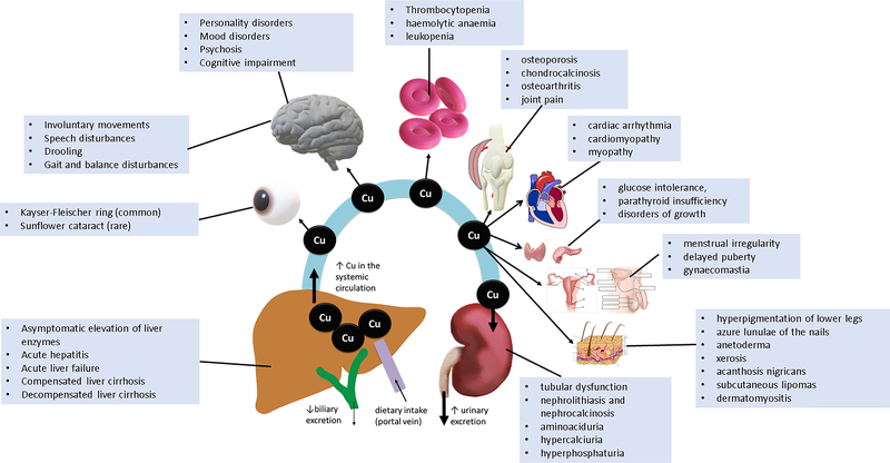
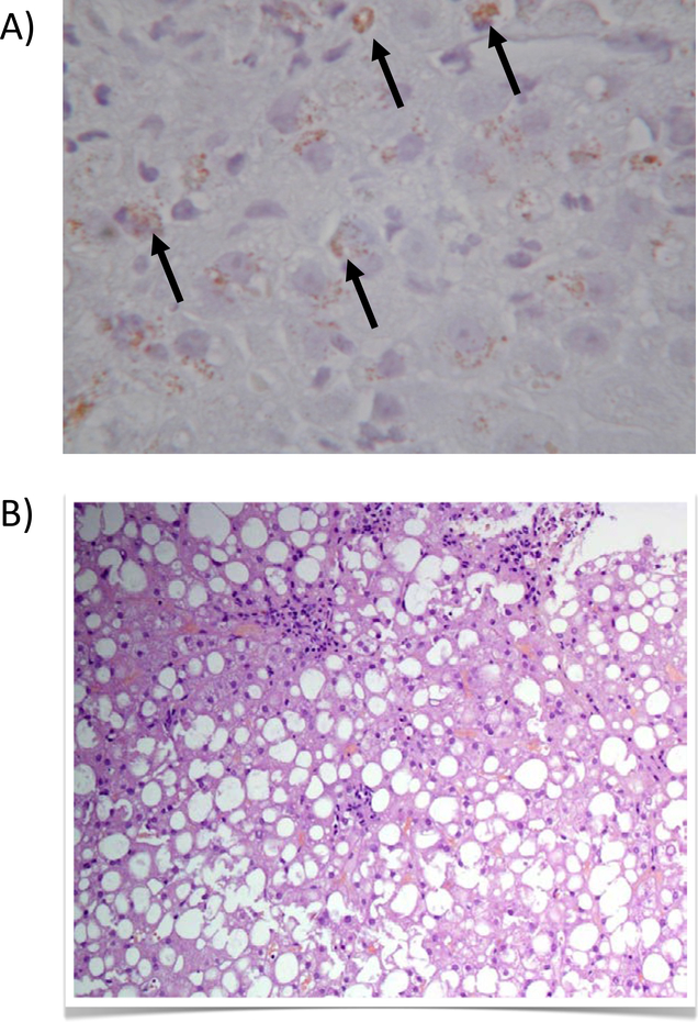
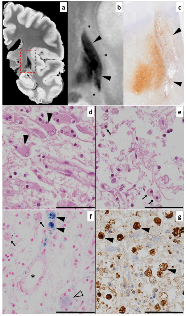
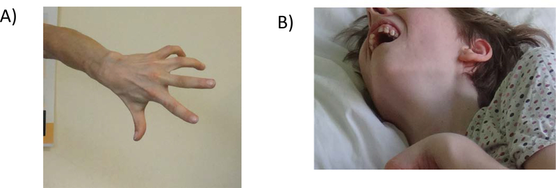
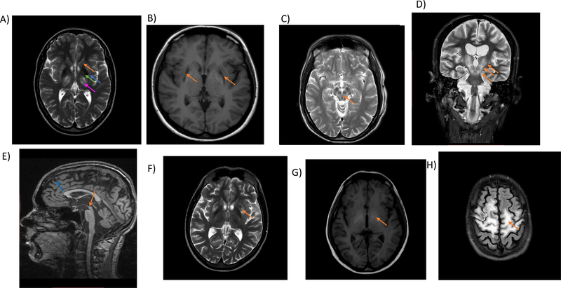
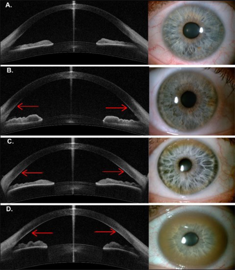
Wilson disease (WD) is a potentially treatable, inherited disorder of copper metabolism that is characterized by the pathological accumulation of copper. WD is caused by mutations in ATP7B, which encodes a transmembrane copper-transporting ATPase, leading to impaired copper homeostasis and copper overload in the liver, brain and other organs. The clinical course of WD can vary in the type and severity of symptoms, but progressive liver disease is a common feature. Patients can also present with neurological disorders and psychiatric symptoms. WD is diagnosed using diagnostic algorithms that incorporate clinical symptoms and signs, measures of copper metabolism and DNA analysis of ATP7B. Available treatments include chelation therapy and zinc salts, which reverse copper overload by different mechanisms. Additionally, liver transplantation is indicated in selected cases. New agents, such as tetrathiomolybdate salts, are currently being investigated in clinical trials, and genetic therapies are being tested in animal models. With early diagnosis and treatment, the prognosis is good; however, an important issue is diagnosing patients before the onset of serious symptoms. Advances in screening for WD may therefore bring earlier diagnosis and improvements for patients with WD.


Conflict of interest statement

Competing interests

A.C. has served on advisory boards for Wilson Therapeutics, Vivet Therapeutics, GMPO and received speaker fees from EVER, Boehringer Ingelheim and Nutricia; P.F. has served on advisory boards for Wilson Therapeutics, Vivet Therapeutics and Univar and received speaker fees from Univar; V.M. has served as a consultant for Kadmon; K.H.W. is on speakers bureau of Abbvie, Alexion, Bayer, BMS, Chiesi, GMPO, Norgine, Novartis, Univar, Wilson Therapeutics, Vivet Therapeutics and received grants (to the institution) from Alexion, Bayer, BMS, Eisai, GMPO, Novartis, Univar and Wilson Therapeutics; M.L.S. has served on advisory boards for Wilson Therapeutics, Vivet Therapeutics, GMPO and Kadmon, is a speaker for Gilead and is on the Medical Advisory Committee of the Wilson disease association; T.L., P.D., S.L. and J.K.R. declare no competing interests.

Figures

Figure 1.
Figure 1.. A timeline of key discoveries in WD
Samuel Alexander Kinnier Wilson described the Wilson disease (WD) in 12 patients in 1912. However, the first cases of WD with dominant tremor symptoms were described in 1883 by Westphal, while the corneal rings pathognomonic of WD were described by Kayser and by Fleischer in 1902–1903. Then followed the discovery of disturbances in copper metabolism as the aetiology of WD and the autosomal recessive inheritance. In 1945, the first treatment for copper toxicosis in WD, British anti-Lewisite (BAL), was introduced. In 1956, d-penicillamine became the first oral drug for WD, which was followed by the availability of zinc salts in 1961 and trientine in 1982. Liver transplantation, as an ultimate treatment for WD, was performed by Starzl and coworkers in 1971. In 1993, the WD gene, ATP7B, was located to chromosome 13q and found to code a P-type ATPase involved in copper transport.
Figure 2.
Figure 2.. Copper homeostasis in the hepatocytes
Cellular copper (Cu) uptake in hepatocytes and other cells is primarily mediated by copper transporter 1, CTR1. A yet unknown cuprireductase and/or extracellular ascorbate provide the reduced copper species for uptake by CTR1. Specialised chaperons shuttle copper to its specific cellular targets: the copper chaperon for superoxide dismutase (CCS) to superoxide dismutase 1 (SOD1), Cox17 to SCO1/2 for subsequent incorporation into cytochrome c oxidase, and antioxidant protein 1 (ATOX1) shuttles copper to the copper transporting ATPases, ATP7A and ATP7B, in the trans-Golgi network (TGN). In the TGN, ATP7B activates ceruloplasmin (Cp) by packaging six copper molecules into apoceruloplasmin, which is then secreted into the plasma. In the cytoplasm, ATP7B sequesters excess copper into vesicles and excretes it across the apical membrane into bile.In the liver, ATP7B provides copper for incorporation into ceruloplasmin (Cp) and is also required for biliary copper excretion. Pathways altered in WD are marked by dashed lines. With reduced/absent levels of ATP7B in WD, there is reduced biliary copper excretion and reduced incorporation of copper into ceruloplasmin. (ref for text content: Scheiber 2017) Image provided by Petr Dusek and Valentina Medici. GSH, glutathione; MT, metallothioneins
Figure 3.
Figure 3.. Copper toxicity in the pathogenesis of Wilson disease
Dietary copper (Cu) is transported via portal vein and sequestered in liver which is the central organ for systemic copper balance. Impairment of biliary copper excretion in Wilson disease caused by ATP7B dysfunction leads to gradual copper accumulation in liver. When the liver capacity to store copper is exhausted, excessive quantities of non-ceruloplasmin-bound copper enter systemic circulation and is deposited in various organs exerting extrahepatic copper toxicity. Copper accumulates in the cornea, brain, red blood cells, skeletal and cardiac muscle cells, synovial membranes of large joints, and renal parenchyma. Non-ceruloplasmin-bound plasma copper is filtrated by renal tubular epithelium and is excreted via urine.
Figure 4.
Figure 4.. Liver pathology of WD
The histochemical demonstration of hepatic copper can be observed as rhodanine-positive granules (part a, black arrows). Early characteristic alterations of the liver pathology in WD include steatosis (part b), which is sometimes indistinguishable from non-alcoholic fatty liver disease. Image b) has been adapted from REF. [Image a) was provided by Professor Ferenci as an original image]
Figure 5.
Figure 5.. Post mortem MRI and histopathology in neurological Wilson disease
a) T2*-weighted post mortem MRI acquired at 7T scanner; b) magnification of the basal ganglia region marked by dashed red rectangle in a), note shrunken and markedly hypointense putamen and globus pallidus (marked by black arrowheads) surrounded by mildly hyperintense area (marked by asterisks); c) low power magnification of Turnbull iron staining displaying corresponding area with MRI on image b), iron staining corresponds with low MRI T2* signal; d & e) Hematoxylin-eosin staining showing reactive astrocytes with large pale nuclei (black arrowheads) and severely damaged tissue with macrophages (black arrows), note that d) corresponds to T2* hyperintense area directly adjacent to putamen while e) corresponds to rarefied area in central putamen; f) Berlin-blue staining showing iron-negative (black arrows) and iron-positive (black arrowheads) macrophages, iron is faintly present also in astrocytes (empty arrowhead) and as iron dust with dominant perivascular distribution (vessel marked by asterisk); g) Ferritin staining shows numerous strongly positive macrophages (black arrowheads) which drive the MRI T2* signal drop. Scale bars in images D-G represent 50 μm.
Figure 6.
Figure 6.. Dystonia, a characteristic symptom in WD
Dystonia is present in at least a third of all patients with a neurological presentation of WD and can be generalised, segmental, multifocal, or focal (part a; focal hand dystonia). The most characteristic WD dystonic presentation is abnormal facial expression or risus sardonicus, which presents as a fixed smile due to dystonia of the risorius muscle (part b, severe dystonia).
Figure 7.
Figure 7.. Brain MRI changes in WD.
Usual abnormalities in brain MRI in patients with WD include symmetric hyperintense changes visualised in T2-weighted images of the basal ganglia, particularly the putamen (blue arrow), caudate nuclei (orange arrow), thalami (pink arrow) and globi pallidi (green arrow)<sup>,,114–117</sup>(part a). In more advanced cases, severe tissue damage can be visualised in T1-weighted images as hypointensity in the putamen (part a, blue arrow; part b, orange arrows). The most spectacular WD changes are described as the ‘face of the giant panda’ in the midbrain (part c). Another common MRI abnormality is increased T2 signal along the dentato-rubro-thalamic pathway (part d), which is a major efferent pathway from the cerebellum involved in movement disorder symptoms, including ataxia, tremor and dystonia. Particularly in severe cases of WD, diffuse brain atrophy (part e) can be seen in the midbrain (orange arrow) and cortex (blue arrow). In brain MRI scans from a 21-year old male with severe liver failure and discrete neurological signs, hyperintense changes in putamina can be visualised in T2-weighted images, which is characteristic of early stages of brain involvement (part f). Hyperintense changes in the globi palidi in T1-weighted images in the same patient are presumably due to manganese accumulation, which is characteristic of hepatic encephalopathy and hypointense changes due to neurodegeneration in the putamina (part g). Very rarely, diffuse white matter changes in both brain hemispheres are observed with preservation of the cortex, probably due to myelin destruction (part h). MRI, magnetic resonance imaging; WD, Wilson disease.
Figure 8.
Figure 8.. Kayser-Fleischer rings visualised by anterior segment optical coherent tomography.
In some patients and in healthy individuals, corneal copper deposits are not seen (part a). However, on other WD patients, copper deposits can be visualised as hyperreflective points by anterior segment optical coherence tomography that are either discrete (part b), on the superior and inferior part of the cornea (part c) or form a complete Kayser-Fleischer ring (part d). Kindly provided by Dr Karina Broniek and Professor Jacek Szaflik from the Department of Ophthalmology, Medical University of Warsaw, Poland

References

    1. Bandmann O, Weiss KH & Kaler SG Wilson’s disease and other neurological copper disorders. Lancet Neurol. 14, 103–113 (2015). - PMC - PubMed
    1. Ferenci P Regional distribution of mutations of the ATP7B gene in patients with Wilson disease: impact on genetic testing. Hum Genet 120, 151–159 (2006). - PubMed
    1. Dzieżyc K, Karliński M, Litwin T & Członkowska A Compliant treatment with anti-copper agents prevents clinically overt Wilson’s disease in pre-symptomatic patients. Eur. J. Neurol 21, 332–337 (2013). - PubMed
    1. EASL Clinical Practice Guidelines: Wilson’s disease. J. Hepatol 56, 671–685 (2012). - PubMed
    1. Roberts EA & Schilsky ML Diagnosis and treatment of Wilson disease: An update. Hepatology 47, 2089–2111 (2008). - PubMed

Publication types

MeSH terms