. 2018 Sep;13(9):1958-1978.
doi: 10.1038/s41596-018-0025-6.

PhIP-Seq characterization of serum antibodies using oligonucleotide-encoded peptidomes

Affiliations

PhIP-Seq characterization of serum antibodies using oligonucleotide-encoded peptidomes

Divya Mohan et al. Nat Protoc. 2018 Sep.

Erratum in

Abstract

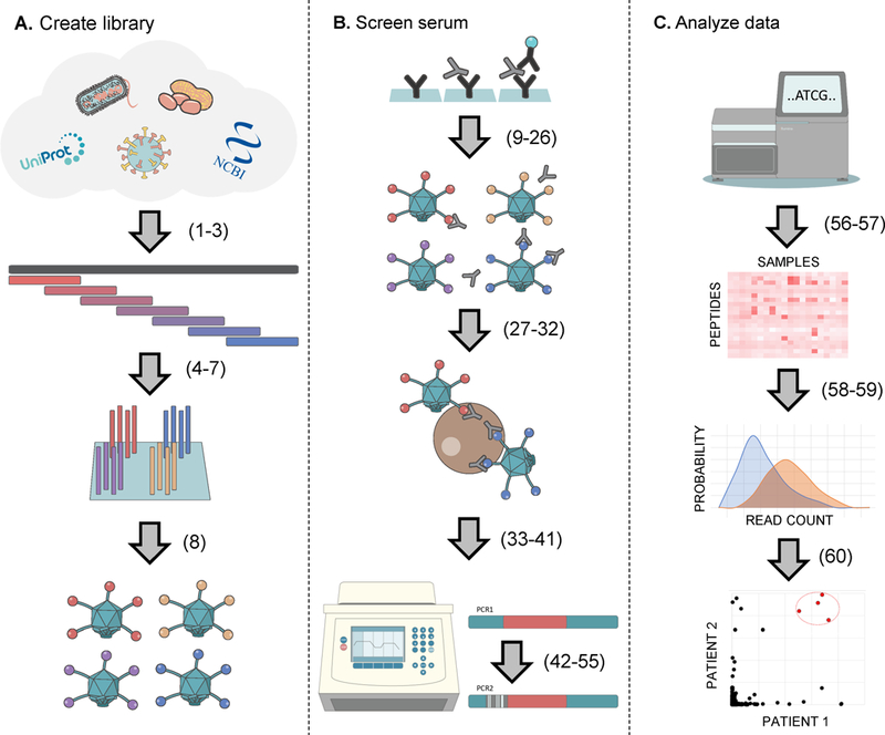
The binding specificities of an individual's antibody repertoire contain a wealth of biological information. They harbor evidence of environmental exposures, allergies, ongoing or emerging autoimmune disease processes, and responses to immunomodulatory therapies, for example. Highly multiplexed methods to comprehensively interrogate antibody-binding specificities have therefore emerged in recent years as important molecular tools. Here, we provide a detailed protocol for performing 'phage immunoprecipitation sequencing' (PhIP-Seq), which is a powerful method for analyzing antibody-repertoire binding specificities with high throughput and at low cost. The methodology uses oligonucleotide library synthesis (OLS) to encode proteomic-scale peptide libraries for display on bacteriophage. These libraries are then immunoprecipitated, using an individual's antibodies, for subsequent analysis by high-throughput DNA sequencing. We have used PhIP-Seq to identify novel self-antigens associated with autoimmune disease, to characterize the self-reactivity of broadly neutralizing HIV antibodies, and in a large international cross-sectional study of exposure to hundreds of human viruses. Compared with alternative array-based techniques, PhIP-Seq is far more scalable in terms of sample throughput and cost per analysis. Cloning and expression of recombinant proteins are not required (versus protein microarrays), and peptide lengths are limited only by DNA synthesis chemistry (up to 90-aa (amino acid) peptides versus the typical 8- to 12-aa length limit of synthetic peptide arrays). Compared with protein microarrays, however, PhIP-Seq libraries lack discontinuous epitopes and post-translational modifications. To increase the accessibility of PhIP-Seq, we provide detailed instructions for the design of phage-displayed peptidome libraries, their immunoprecipitation using serum antibodies, deep sequencing-based measurement of peptide abundances, and statistical determination of peptide enrichments that reflect antibody-peptide interactions. Once a library has been constructed, PhIP-Seq data can be obtained for analysis within a week.


Figures

Figure 1.
Figure 1.
Overview of the PhIP-seq methodology. Procedure step numbers are indicated in parentheses. A. A protein database is downloaded or designed. The pepsyn software is used to tile the protein sequences with overlapping peptide sequences. The oligonucleotide library encoding the peptide sequences is synthesized. The oligonucleotide library is PCR amplified with adapters for cloning into the phage display vector of choice. B. ELISA is used to quantify each sample’s IgG content for normalizing amount of antibody input into each phage binding reaction. Antibodies and their bound phage are captured using protein A/G coated magnetic beads. The library of peptide encoding DNA sequences are amplified by PCR directly from the immunoprecipitate. A second round of hemi-nested PCR is used to add sample-specific barcodes and sequencing adapters to the PCR1 product. Barcoded amplicons are pooled for sequencing on an Illumina instrument. C. Fastq sequencing files are demultiplexed and aligned to the reference sequences to obtain a count matrix. Statistical analysis of the count matrix is performed to determine peptide enrichments. Project specific analysis of peptide enrichments (e.g. identification of a common autoantigen) can then be carried out.
Figure 2.
Figure 2.
Bioinformatics workflows. Procedure step numbers are indicated. A. Pepsyn workflow. Workflow for designing a peptide library. We only provide an outline of the protocol as this stage will likely be customized depending on your library/preferences. B. PhIP-stat workflow for PhIP-Seq data analysis.
Figure 3.
Figure 3.
Primer-depleted, pooled VirScan PCR2 products run at a higher molecular weight than expected (shown on a 2% agarose gel in lithium borate). Lane 1: 1Kb Plus DNA Ladder (Invitrogen) Lane 2: empty Lane 3: Primer-depleted VirScan PCR2 product. Lane 4: Product of VirScan PCR3, without replenishment of primers. Lane 5: Product of VirScan PCR3, with replenishment of primers of primers (P5 and P7.2).
Figure 4.
Figure 4.
Organization of bacteriophage genome, primer binding sites and PCR products. The peptide coding sequence, originally derived from the oligonucleotide library, is cloned into the T7 genome as a C-terminal fusion with the 10B capsid protein. PCR1 (steps 42–44): T7-Pep2_PCR1_F is used as the outside PCR1 primer. T7-Pep2_PCR1_R+ad_min is used as the reverse PCR1 primer, and to add the minimal adapter required for subsequent addition of the sample barcode during PCR2. PCR2 (steps 45–49): The product of PCR1 is used as the template for PCR2. T7-Pep2_PCR2_F_P5 is used as the forward PCR2 primer, and to add the required Illumina P5 adapter (and optionally the i5 dual index, not shown). The set of primers called ad_min_BCX_P7 (where X defines the sample-specific DNA barcode) are used individually as the reverse PCR2 primers, to add the sample-specific DNA barcode, and to add the required Illumina P7 adapter. After pooling PCR2 products from all the samples, a single round of PCR3 is performed (steps 52–53), using the P5 and P7.2 primers, which ensures the DNA libraries are fully double-stranded. Illumina sequencing (step 55): T7-VirScan_SP is the Read 1 sequencing primer used to obtain the peptide coding sequence. Index_SP is the standard Illumina Multiplex Single End Read 2 sequencing primer used to obtain the sample-specific barcode. The sequences generated from the Illumina sequencing run are shown as dashed lines: Read 1 obtains the first 50 bases of the peptide coding sequence and Read 2 is the 8 cycle index read
Figure 5.
Figure 5.
Output from the sequencing data analysis pipeline. (A) Sjögren’s Syndrome (SS) patient A’s serum sample was screened against the human peptidome library and analyzed in duplicate. Read counts were divided by the total reads, multiplied by 1×10⁶ and then plotted. The scatter plot illustrates the reproducibility of the post-immunoprecipitation clonal distributions between the two replicas. Red filled circles highlight peptides from the Ro52 (TRIM21) protein, to which this patient was known to have autoantibodies. (B) Comparison of patient A’s immunoprecipitated clonal distribution to that of a set of mock IPs (no sample input), which illustrates (i) the bias in the starting library and (ii) antibody-dependent enrichment of specific phage clones (including strong enrichment of three Ro52 peptides). (C) Generalized Poisson (GP) based p-values calculated using the data in (A) as input. Background bias has been removed from this distribution, which illustrates reproducible antibody dependent enrichments. (D) Comparing the enrichment scores (-log10 p-values) of two different individuals illustrates their largely non-overlapping enrichment profiles. However, three peptides from Ro52 are among the shared enrichments. De-identified serum samples were analyzed in accordance with JHU Human Subject Research exemption IRB00049327.

References

    1. Larman HB et al. Autoantigen discovery with a synthetic human peptidome. Nature biotechnology 29, 535–541 (2011). - PMC - PubMed
    1. Larman HB et al. PhIP-Seq characterization of autoantibodies from patients with multiple sclerosis, type 1 diabetes and rheumatoid arthritis. Journal of autoimmunity 43, 1–9 (2013). - PMC - PubMed
    1. Larman HB et al. Cytosolic 5’-nucleotidase 1A autoimmunity in sporadic inclusion body myositis. Annals of neurology 73, 408–418 (2013). - PubMed
    1. Finton KA et al. Ontogeny of recognition specificity and functionality for the broadly neutralizing anti-HIV antibody 4E10. PLoS pathogens 10, e1004403 (2014). - PMC - PubMed
    1. Xu GJ et al. Viral immunology. Comprehensive serological profiling of human populations using a synthetic human virome. Science 348, aaa0698 (2015). - PMC - PubMed

Publication types