. 2018 Aug 23:9:1190.
doi: 10.3389/fphys.2018.01190. eCollection 2018.

NFAT5 Isoform C Controls Biomechanical Stress Responses of Vascular Smooth Muscle Cells

Affiliations

NFAT5 Isoform C Controls Biomechanical Stress Responses of Vascular Smooth Muscle Cells

Maren Zappe et al. Front Physiol. .

Abstract

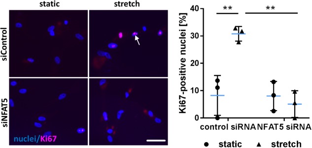
Vascular cells are continuously exposed to mechanical stress that may wreak havoc if exceeding physiological levels. Consequently, mechanisms facing such a challenge are indispensable and contribute to the adaptation of the cellular phenotype. To this end, vascular smooth muscle cells (VSMCs) activate mechanoresponsive transcription factors promoting their proliferation and migration to initiate remodeling the arterial wall. In mechanostimulated VSMCs, we identified nuclear factor of activated T-cells 5 (NFAT5) as transcriptional regulator protein and intended to unravel mechanisms controlling its expression and nuclear translocation. In cultured human VSMCs, blocking RNA synthesis diminished both baseline and stretch-induced NFAT5 mRNA expression while inhibition of the proteasome promoted accumulation of the NFAT5 protein. Detailed PCR analyses indicated a decrease in expression of NFAT5 isoform A and an increase in isoform C in mechanoactivated VSMCs. Upon overexpression, only NFAT5c was capable to enter the nucleus in control- and stretch-stimulated VSMCs. As evidenced by analyses of NFAT5c mutants, nuclear translocation required palmitoylation, phosphorylation at Y143 and was inhibited by phosphorylation at S1197. On the functional level, overexpression of NFAT5c forces its accumulation in the nucleus as well as transcriptional activity and stimulated VSMC proliferation and migration. These findings suggest that NFAT5 is continuously expressed and degraded in resting VSMCs while expression and accumulation of isoform C in the nucleus is facilitated during biomechanical stress to promote an activated VSMC phenotype.

Keywords: NFAT5; TonEBP; hypertension; transcriptional regulation; vascular smooth muscle cells; wall stress.


Figures

FIGURE 1
FIGURE 1
Analysis of NFAT5 mRNA expression in VSMCs. Localization of NFAT5 protein was detected by immunofluorescence (A, scale bar: 100 μm) and NFAT5 mRNA expression was analyzed by semi-quantitative PCR (B, p < 0.05, n = 3, unpaired, 2-tailed Student’s t-test; the expression of the housekeeping gene RPL32 served as internal standard) in resting and stretch-stimulated (24 h) cultured HUASMCs. After treatment (24 h) with actinomycin-D NFAT5 mRNA expression declined (C, ActD; ∗∗∗p < 0.001, ∗∗p < 0.01, n = 4; one-way ANOVA and Tukey post-test).
FIGURE 2
FIGURE 2
Analysis of NFAT5 protein abundance upon inhibition of the proteasome. HUASMCs were treated (24 h) with increasing concentrations of bortezomib and NFAT5 was detected by immunostaining. The percentage of NFAT5-positive nuclei were quantified by automated image analysis (A, ∗∗∗p < 0.001, n = 3, one-way ANOVA and Tukey post-test; scale bar 100 μm). NFAT5 protein abundance was determined after treatment with 10 nM bortezomib (24 h) in nuclear (B) and cytosolic (C) fractions of cell lysates (B,C, ∗∗∗p < 0.001/∗∗p < 0.01 vs. DMSO, n = 3, unpaired, 2-tailed Student’s t-test; histone H3 and tubulin served as loading controls).
FIGURE 3
FIGURE 3
Biomechanical stretch induces NFAT5c translocation into the nucleus. Expression of NFAT5 isoforms after stretch was quantitated by semi-quantitative PCR (A, p < 0.05 vs. control, n = 3, unpaired, 2-tailed Student’s t-test, n.s.: not significant). DDK-specific immunostaining was used for detection of NFAT5 isoform localization in HUASMCs exposed to biomechanical stretch for 24 h, white arrows indicate localization of the nuclei, scale bar: 50 μm). Automated quantification of DDK-positive nuclei in transfected cells 24 h after stretch (B, ∗∗∗p < 0.001, p < 0.05, n = 3, one-way ANOVA and Tukey post-test).
FIGURE 4
FIGURE 4
Posttranslational modifications are required for NFAT5c to enter the nucleus of mechanoactivated VSMCs. NFAT5c was overexpressed in HUASMCs and the cells were exposed to biomechanical stretch (24 h). HUASMCs were treated with 2-bromopalmitate (inhibitor of protein palmitoylation, (A), ∗∗∗p < 0.001, n = 4, one-way ANOVA and Tukey post-test, scale bar: 50 μm). Site directed mutagenesis of NFAT5c at serine 1197 (serine → glutamic acid) was performed to mimic phosphorylation at this site (B, p < 0.05, ∗∗p < 0.01, n = 6–7, one-way ANOVA and Tukey post-test, scale bar: 50 μm). Site directed mutagenesis of NFAT5c at tyrosine 143 (tyrosine → alanine) was performed to prevent phosphorylation at this site (C, p < 0.05, n = 3, one-way ANOVA and Tukey post-test, scale bars: 50 μm). In all approaches (a-c), localization of NFAT5c in the nuclei was determined by immunofluorescence-based detection of DDK (automated image analysis) in stretch-stimulated HUASMCs.
FIGURE 5
FIGURE 5
Analysis of nuclear translocation of NFAT5 upon knockdown of ABL1. HUASMCs were transfected with control (scrambled) siRNA or ABL1-specific siRNA (ABL1 Silencer®Select Validated siRNA, S864, Ambion, Applied Biosystem, Thermo Fisher Scientific, Darmstadt, Germany) which decreased ABL1 expression by ∼50% as evidenced by PCR analyses (A). Transfected HUASMCs were exposed to biomechanical stretch for 24 h. NFAT5-specific immunofluorescence was detected by automated image analysis to assess the percentage of NFAT5-positive nuclei (B, p < 0.05, one-way ANOVA and Tukey post-test; control siRNA n = 5, ABL1 siRNA n = 10). Dasatinib (inhibitor of BCR-ABL kinases, 5 nM) treatment of stretch-stimulated HUASMCs decreased the number of NFAT5 positive nuclei as evidenced by automated immunofluorescence analyses (C, p < 0.05, one-way ANOVA and Tukey post-test, n = 3). Additionally, HUASMCs were treated with Dasatinib (Dasa; 5 nM) or DMSO for 1 h and exposed to biomechanical stretch for 24 h. Accumulation of NFAT5 in the nucleus was determined in nuclear lysates by immunoblot techniques with (nuclear) histone H3 as loading marker (D, ∗∗∗p < 0.05, n.s.: not significant, one-way ANOVA and Tukey post-test, n = 3).
FIGURE 6
FIGURE 6
Overexpression of NFAT5c promotes its nuclear accumulation and activates VSMCs. HUASMCs were transduced with adenovirus to overexpress NFAT5c (Ad-N5) or (Ad-PL) control plasmid, NFAT5-expression was quantitated by qRT-PCR, immunostaining and immunoblot of nuclear and cytosolic lysates 48 h after transfection (A–C, ∗∗p < 0.01 vs. Ad-PL, n = 6, unpaired, 2-tailed Student’s t-test; histone H3 and β-actin served as loading controls). Expression levels of NFAT5 target gene TNC was assessed by qRT-PCR (D, ∗∗p < 0.01 vs. Ad-PL, n = 6, unpaired, 2-tailed Student’s t-test). The migratory capacity of collagen-embedded NFAT5c-overexpressing spheroids was analyzed by assessing the number of sprouts, mean sprout length and cumulative sprout length after 24 h (E, ∗∗∗p < 0.001 vs. Ad-PL, n = 10 spheroids/condition, 1 out of 3 independent experiments with comparable results, unpaired, 2-tailed Student’s t-test; scale bar: 200 μm). Proliferation of NFAT5c-overexpressing HUASMCs 72 h after transfection by immunostaining of the proliferation marker Ki67 (F, p < 0.05, n.s.: not significant, vs. Ad-PL, n = 4, unpaired, 2-tailed Student’s t-test, scale bar: 100 μm).
FIGURE 7
FIGURE 7
Gene set enrichment analyses (GSEA) based on the KEGG pathway database. HUASMCs from three donors were treated with NFAT5-specific or control siRNA and exposed to biomechanical stretch for 24 h. RNA was isolated and processed for genome array analysis (HuGene-1 0-st-v1 array, Affymetrix). The table (A) lists all significantly DOWN-regulated gene sets associated with the indicated pathways/functions (log2-fold regulation, p < 0.05 NFAT5 knockdown vs. control, n = 3, one-way ANOVA; access# for full microarray data via Gene Expression Omnibus (GEO): GSE106274). Exemplary results of the subsequent gene set enrichment analyses (GSEA) for selected gene sets (enrichment plots) and the corresponding statistical values are shown [B–D; NES: normalized enrichment score; p: p-value (ANOVA); FDR: false discovery rate]. Additionally, the heat map corresponding to the enrichment plot for cell cycle-associated genes is shown.
FIGURE 8
FIGURE 8
Knockdown of NFAT5 inhibits stretch-induced proliferation of HUASMCs. HUASMCs were treated with NFAT5-specific or control siRNA and exposed to biomechanical stretch for 24 h. Proliferation was assessed by detecting the proliferation marker Ki67 via immunofluorescence techniques (arrow) and determining the percentage of Ki67-positive nuclei (∗∗p < 0.01, one-way ANOVA and Tukey post-test, n = 3; scale bar: 50 μm).

References

    1. Ahn J. D., Morishita R., Kaneda Y., Lee S. J., Kwon K. Y., Choi S. Y., et al. (2002). Inhibitory effects of novel AP-1 decoy oligodeoxynucleotides on vascular smooth muscle cell proliferation in vitro and neointimal formation in vivo. Circ. Res. 90 1325–1332. 10.1161/01.RES.0000023200.19316.D5 - DOI - PubMed
    1. AlGhatrif M., Lakatta E. G. (2015). The conundrum of arterial stiffness, elevated blood pressure, and aging. Curr. Hypertens. Rep. 17:12. 10.1007/s11906-014-0523-z - DOI - PMC - PubMed
    1. Bergman M. R., Cheng S., Honbo N., Piacentini L., Karliner J. S., Lovett D. H. (2003). A functional activating protein 1 (AP-1) site regulates matrix metalloproteinase 2 (MMP-2) transcription by cardiac cells through interactions with JunB-Fra1 and JunB-FosB heterodimers. Biochem. J. 369 485–496. 10.1042/BJ20020707 - DOI - PMC - PubMed
    1. Dahl S. C., Handler J. S., Kwon H. M. (2001). Hypertonicity-induced phosphorylation and nuclear localization of the transcription factor TonEBP. Am. J. Physiol. Cell Physiol. 280 C248–C253. 10.1152/ajpcell.2001.280.2.C248 - DOI - PubMed
    1. Dalski A., Schwinger E., Zuhlke C. (2001). Genomic organization of the human NFAT5 gene: exon-intron structure of the 14-kb transcript and CpG-island analysis of the promoter region. Cytogenet. Cell Genet. 93 239–241. 10.1159/000056990 - DOI - PubMed