Review
. 2014 Aug;1(1):57-74.
doi: 10.2217/mmt.14.10. Epub 2014 Sep 5.

Melanoma and nuclear medicine

Affiliations
Review

Melanoma and nuclear medicine

Andrés Perissinotti et al. Melanoma Manag. 2014 Aug.

Abstract

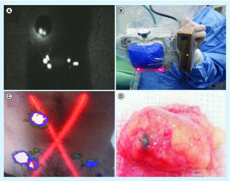
Supported by a large body of published work, the contribution of nuclear medicine technologies to the assessment of melanoma has been increasing in recent years. Lymphoscintigraphy-assisted sentinel lymph node biopsy and PET are in continuous evolution with the aid of technological imaging advances, making it possible to fuse functional and anatomic images (e.g., with SPECT/CT, PET/CT and 3D rendering systems). The development of hybrid fluorescent-radioactive tracers that enable high-quality preoperative lymphoscintigraphy and SPECT/CT, and the optimization of modern intraoperative portable imaging technologies, such as free-hand SPECT and portable γ-cameras, are important innovations that have improved sentinel lymph node identification in complex anatomical areas, such as the pelvis and head and neck. Concurrently, 18F-fluorodeoxyglucose-PET has proved its usefulness in the clinical staging and treatment decision-making process, and there is also emerging evidence regarding its utility in the evaluation of therapeutic response. The potential uses of other novel PET radiotracers could open up a new field of use for this technique. In this article, we review the current and future role of nuclear medicine in the management of melanoma.

Keywords: PET/CT; fluorescence; lymph node biopsy; melanoma; portable imaging technologies; radio-guided surgery.


Conflict of interest statement

Financial & competing interests disclosure The authors have no relevant affiliations or financial involvement with any organization or entity with a financial interest in or financial conflict with the subject matter or materials discussed in the manuscript. This includes employment, consultancies, honoraria, stock ownership or options, expert testimony, grants or patents received or pending, or royalties. No writing assistance was utilized in the production of this manuscript.

Figures

<b>Figure 1.</b>
Figure 1.. Patient with a melanoma in the right lumbar region.
(A) Delayed (2 h) planar image showing drainage to both sides of the groin. (B) Hybrid portable γ-camera equipped with an optical device. (C) Fused image obtained with the hybrid γ-camera showing the same distribution as in (A), but with an anatomical overlay. (D) Sentinel node excised from the right groin, showing a black metastatic deposit.
<b>Figure 2.</b>
Figure 2.. Detection of sentinel nodes using free-hand SPECT navigation.
(A) Planar lymphoscintigraphy in a patient with a right malar melanoma showing two sentinel nodes in the cervical area. (B) 3D volume-rendered image based on SPECT/CT data showing these sentinel nodes to be in cervical levels Ib and IIa. (C) Intraoperative free-hand SPECT navigation. (D) Excision of one of the two sentinel nodes.
<b>Figure 3.</b>
Figure 3.. Nanocolloid and Indocyanine Green hybrid radiotracer approach.
(A & B) After the addition of Indocyanine Green to nanocolloid, different doses are prepared for administration in a similar way to the radiotracer-only approach. (C) Preoperative lymphoscintigraphy is performed as with conventional radiotracer-only approaches. (D) Intraoperative imaging with a dedicated fluorescence camera shows a high-resolution image of a sentinel node on the screen.
<b>Figure 4.</b>
Figure 4.. Patient with a melanoma on the dorsum of the right foot.
(A) The planar posterior view shows two lymphatic channels. One goes up to the groin and the other to the popliteal fossa where two nodes are depicted, while there is a hint of a third node downstream. (B) The 3D volume-rendered reconstructed image provides the anatomical habitat of the two nodes and confirms the presence of the faint third node. (C) The intraoperative image with a portable γ-camera shows the first two popliteal nodes. SPECT/CT is a very useful tool for planning the surgical approach. (D) In this case, a very deep popliteal node is demonstrated in the axial slice, requiring the surgeon to make a long incision.
<b>Figure 5.</b>
Figure 5.. Sentinel nodes in unexpected areas of lymphatic drainage.
(A) Anterior planar image of a patient with a melanoma in the right flank showing not only drainage to the right groin, but also to the medial area of the trunk (arrow). Note that the sentinel node is clearly defined on (B & C) 3D imaging and (D) transversal SPECT/CT in the paravertebral muscle on the right. (E) In another patient with a melanoma of the back, posterior planar imaging shows drainage to both axillae and the left supraclavicular region (arrow). This sentinel node is anatomically indicated on (F) 3D imaging and transversal (G) SPECT/CT and corresponds on (H) CT with a slightly enlarged node (circle), which contained metastases at histopathology. (I) In another patient with a melanoma of the left underarm, anterior planar imaging shows drainage to the left axilla and to the area of the left elbow (arrow). (J–L) On SPECT/CT, this sentinel node is seen as a dorsal subcutaneous node.
<b>Figure 6.</b>
Figure 6.. Radio-guided surgery of isolated melanoma metastases.
(A) In a melanoma patient, PET/CT showed a solitary 18F-fluorodeoxyglucose-avid lesion in the right breast. (B) After ultrasound-guided injection of a radioactive tracer, the lesion was indicated by (C & D) SPECT/CT and subsequently (E) resected by the surgeon under guidance of a portable γ-ray device.
<b>Figure 7.</b>
Figure 7.. Widespread disease.
(A & B) PET and PET/CT fused images showing extensive 18F-fluorodeoxyglucose deposits throughout the body, especially in the liver and bone. (C) PET/CT offers an overview of the lesions in the 3D reconstruction display.

References

    1. WHO Ultraviolet radiation and the INTERSUN Programme. http://www.who.int/uv/faq/skincancer/en/index1.html
    1. Balch CM, Soong SJ, Gershenwald JE, et al. Prognostic factors analysis of 17,600 melanoma patients: validation of the American Joint Committee on Cancer melanoma staging system. J. Clin. Oncol. 2001;19(16):3622–3634. - PubMed
    1. Balch CM, Gershenwald JE, Soong S-J, et al. Final version of 2009 AJCC melanoma staging and classification. J. Clin. Oncol. 2009;27(36):6199–6206. - PMC - PubMed
    1. Mohr P, Eggermont AMM, Hauschild A, Buzaid A. Staging of cutaneous melanoma. Ann. Oncol. 2009;20(Suppl. 6):vi14–vi21. - PMC - PubMed
    1. Testori A, De Salvo GL, Montesco MC, et al. Clinical considerations on sentinel node biopsy in melanoma from an Italian multicentric study on 1,313 patients (SOLISM-IMI) Ann. Surg. Oncol. 2009;16(7):2018–2027. - PubMed

LinkOut - more resources