Review
. 2018 Jun 28;5(1):MMT06.
doi: 10.2217/mmt-2017-0022. eCollection 2018 Jun.

Melanoma & nuclear medicine: new insights & advances

Affiliations
Review

Melanoma & nuclear medicine: new insights & advances

Andrés Perissinotti et al. Melanoma Manag. .

Abstract

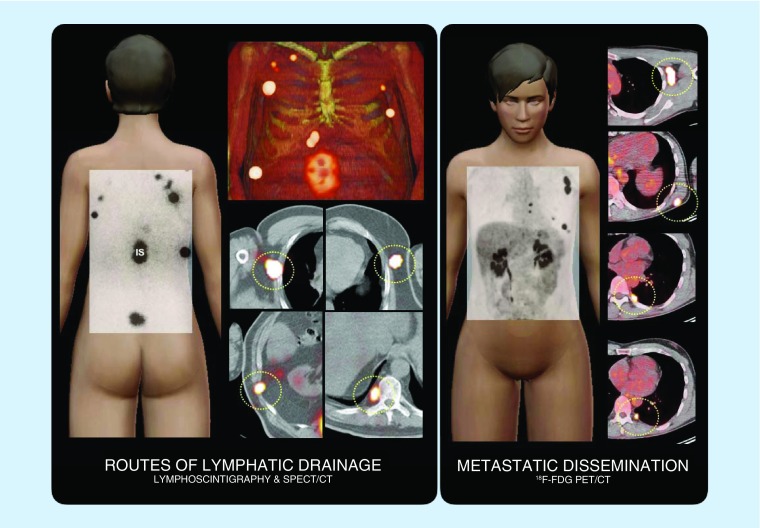
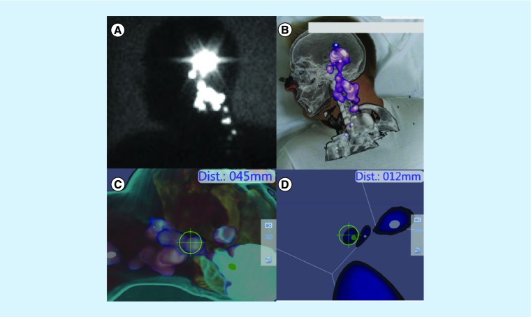
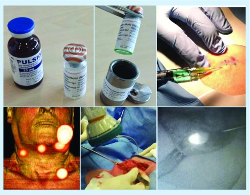
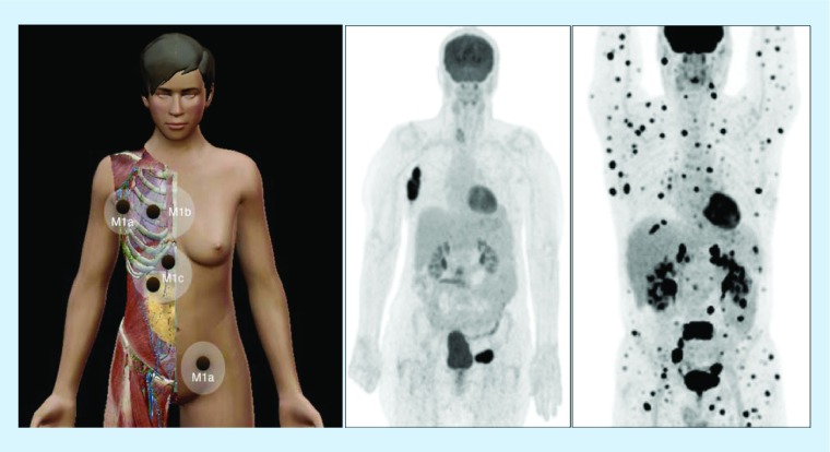
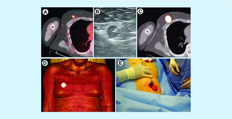
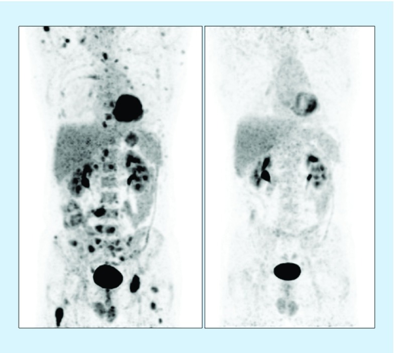
The contribution of nuclear medicine to management of melanoma patients is increasing. In intermediate-thickness N0 melanomas, lymphoscintigraphy provides a roadmap for sentinel node biopsy. With the introduction of single-photon emission computed tomography images with integrated computed tomography (SPECT/CT), 3D anatomic environments for accurate surgical planning are now possible. Sentinel node identification in intricate anatomical areas (pelvic cavity, head/neck) has been improved using hybrid radioactive/fluorescent tracers, preoperative lymphoscintigraphy and SPECT/CT together with modern intraoperative portable imaging technologies for surgical navigation (free-hand SPECT, portable gamma cameras). Furthermore, PET/CT today provides 3D roadmaps to resect 18F-fluorodeoxyglucose-avid melanoma lesions. Simultaneously, in advanced-stage melanoma and recurrences, 18F-fluorodeoxyglucose-PET/CT is useful in clinical staging and treatment decision as well as in the evaluation of therapy response. In this article, we review new insights and recent nuclear medicine advances in the management of melanoma patients.

Keywords: 18F-FDG-PET/CT; SPECT/CT; intraoperative technologies; radioguided surgery; sentinel node biopsy.


Conflict of interest statement

Financial & competing interests disclosure The authors have no relevant affiliations or financial involvement with any organization or entity with a financial interest in or financial conflict with the subject matter or materials discussed in the manuscript. This includes employment, consultancies, honoraria, stock ownership or options, expert testimony, grants or patents received or pending, or royalties. No writing assistance was utilized in the production of this manuscript.

Figures

<b>Figure 1.</b>
Figure 1.. Routes of lymphatic drainage and metastatic dissemination.
On the left, schematic illustration of a case showing lymphatic drainage from the injections (IS) around the site of the primary melanoma on the back to intercostal, axillary and paravertebral (circles) sentinel lymph nodes as assessed by lymphoscintigraphy and SPECT/CT. On the right, another case where PET/CT shows FDG-avid lymph node metastases in left axilla, left scapular area and left paravertebral (circles). FDG: 18F-fluorodeoxyglucose; IS: Injection site; SPECT/CT: Single-photon emission computed tomography images with integrated computed tomography.
<b>Figure 2.</b>
Figure 2.. Patient with a melanoma on the dorsum of the right foot.
(A) The planar posterior view shows two lymphatic channels. One goes up to the groin and the other to the popliteal fossa where two nodes are depicted, while there is a hint of a third node downstream. (B) The 3D volume-rendered reconstructed image provides the anatomical habitat of the two nodes and confirms the presence of the faint third node. (C) The intraoperative image with a portable gamma camera shows the first two popliteal nodes. SPECT/CT is a very useful tool for planning the surgical approach. (D) In this case, a very deep popliteal node is demonstrated in the axial slice, requiring the surgeon to make a long incision. SPECT/CT: Single-photon emission computed tomography images with integrated computed tomography. Reproduced with permission from [1] © Future Medicine Ltd. (2014).
<b>Figure 3.</b>
Figure 3.. Sentinel nodes in unexpected areas of lymphatic drainage.
(A) Anterior planar image of a patient with a melanoma in the right flank showing not only drainage to the right groin but also to the medial area of the trunk (arrow). Note that the sentinel node is clearly defined on (B & C) 3D imaging and (D) transversal SPECT/CT in the paravertebral muscle on the right. (E) In another patient with a melanoma of the back, posterior planar imaging shows drainage to both axillae and the left extracurricular region (arrow). This sentinel node is anatomically indicated on (F) 3D imaging and transversal (G) SPECT/CT and corresponds on (H) CT with a slightly enlarged node (circle), which contained metastases at histopathology. (I) In another patient with a melanoma of the left underarm, anterior planar imaging shows drainage to the left axilla and to the area of the left elbow (arrow). (J–L) On SPECT/CT, this sentinel node is seen as a dorsal subcutaneous node. SPECT/CT: Single-photon emission computed tomography images with integrated computed tomography. Reproduced with permission from [1] © Future Medicine Ltd. (2014).
<b>Figure 4.</b>
Figure 4.. Patient with frontal melanoma.
Fourty-eight-year-old female with a left-sided frontal melanoma (2.5 mm Breslow thickness). Lateral view planar image showed several lymphatic channels aiding to different SNs (A). SPECT/CT data with volume rendering showed a preauricular SN and other SNs in left cervical level II situation (B). Portable gamma camera demonstrated significant activity in those areas but without any anatomical environment (C). Hybrid gamma camera (with and optical component) provided a more convenient anatomical landscape (D). Placing the hybrid gamma camera at different distances allows the nuclear medicine physician or surgeon to better depict and overview of the SN distribution (E) and to mark very precisely the hot nodes on the skin (F). SN: Sentinel node; SPECT/CT: Single-photon emission computed tomography images with integrated computed tomography.
<b>Figure 5.</b>
Figure 5.. Free-hand single-photon emission computed tomography.
Delayed lateral planar image belongs to a 34-year-old male patient with a melanoma (3.2 mm Breslow thickness) in his left ear. Several hot spots are depicted from preauricular area to left cervical (A). After loading SPECT/CT-tracked data volume, rendering reconstruction clearly depicts the lymph nodes in the anatomical environment of level II–III in the neck (B). Using tracked free-hand SPECT acquisition navigation to the nodes is possible (C) and every node can be observed as a 3D virtual spot. The distance from the tip of the gamma probe (12 mm) is depicted in real-time (D). SPECT/CT: Single-photon emission computed tomography images with integrated computed tomography.
<b>Figure 6.</b>
Figure 6.. Nanocolloid and indocyanine green hybrid radiotracer.
After addition of ICG to radioactive nanocolloid the hybrid tracer ICG-99mTc-nanocolloid is injected intracutaneously around the excision scar of a melanoma (above). On SPECT/CT (below on the left), drainage from the primary lesion in the left cheek to four sentinel lymph nodes in both sides of the neck is observed. Subsequently, a dedicated fluorescence camera (middle) enables intraoperative sentinel lymph node identification and excision (on the right). ICG: Indocyanine green; SPECT/CT: Single-photon emission computed tomography images with integrated computed tomography.
<b>Figure 7.</b>
Figure 7.. Melanoma dissemination.
On the left, scheme illustrating disseminated disease in melanoma with distant metastases in lymph nodes (M1a), lung (M1b) and liver/bowel (M1c). Middle, 18F-FDG-avid lymph node metastases in right axilla and left groin as displayed on PET with maximum intensity projection. On the right, multiple FDG-metastases in another patient with widespread disease. FDG: 18F-fluorodeoxyglucose.
<b>Figure 8.</b>
Figure 8.. Radioguided surgery of isolated melanoma metastases.
(A) In a melanoma patient, PET/CT showed a solitary 18F-FDG-avid lesion in the right breast. (B) After ultrasound-guided injection of a radioactive tracer, the lesion was indicated by (C & D) SPECT/CT and subsequently (E) resected by the surgeon under guidance of a portable gamma-ray device. FDG: 18F-fluorodeoxyglucose; SPECT/CT: Single-photon emission computed tomography images with integrated computed tomography. Reproduced with permission from [1] © Future Medicine Ltd. (2014).
<b>Figure 9.</b>
Figure 9.. Therapy response assessment with PET.
Patient with multiple FDG-avid metastases on baseline PET (on the left) with complete response on PET after therapy with BRAF-inhibitors (on the right). FDG: 18F-fluorodeoxyglucose.

References

    1. Perissinotti A, Vidal-Sicart S, Nieweg O, Olmos RV. Melanoma and nuclear medicine. Melanoma Manag. 2014;1(1):57–74. - PMC - PubMed
    1. Balch CM, Soong SJ, Gershenwald JE, et al. Prognostic factors analysis of 17,600 melanoma patients: validation of the American Joint Committee on Cancer melanoma staging system. J. Clin. Oncol. 2001;19(16):3622–3634. - PubMed
    1. Morton DL, Cochran AJ, Thompson JF, et al. Sentinel node biopsy for early-stage melanoma: accuracy and morbidity in MSLT-I, an international multicenter trial. Ann. Surg. 2005;242(3):302–311. - PMC - PubMed
    2. •• Prospective multicenter study that confirmed the value of early nodal evaluation and treatment of melanoma based on sentinel node biopsy (SNB).

    1. Morton DL, Thompson JF, Cochran AJ, et al. Sentinel-node biopsy or nodal observation in melanoma. N. Engl. J. Med. 2006;355(13):1307–1317. - PubMed
    1. Morton DL, Thompson JF, Cochran AJ, et al. Final trial report of sentinel-node biopsy versus nodal observation in melanoma. N. Engl. J. Med. 2014;370(7):599–609. - PMC - PubMed

LinkOut - more resources