Review
. 2014 Oct;1(4):395-408.
doi: 10.2217/hep.14.22. Epub 2014 Dec 11.

Predicting recurrence following radiofrequency percutaneous ablation for hepatocellular carcinoma

Affiliations
Review

Predicting recurrence following radiofrequency percutaneous ablation for hepatocellular carcinoma

Nathalie Ganne-Carrié et al. Hepat Oncol. 2014 Oct.

Abstract

Within 5 years after percutaneous ablation of hepatocellular carcinoma, roughly 70% of patients experience tumor recurrence. Relapses beyond curative options affected patients' survival. Ablation shares with resection common predictive factors of recurrence as size of the tumor, multinodularity and presence of vascular invasion. High serum α-fetoprotein level and markers of severity of underlying liver disease have also been found to be associated with recurrence and even survival. However, predictive values for recurrence of technical factors, histopathological and molecular tumors' features have been rarely studied. Few comparative studies have shown that ablation techniques impact recurrence rates. Moreover, although ablation does not allow analysis of the whole tumor, some reports suggest that biopsies allow histopathological and even molecular testing of the risk of recurrence.

Keywords: cirrhosis; hepatocellular carcinoma; percutaneous ablation; radiofrequency ablation; recurrence.


Conflict of interest statement

Financial & competing interests disclosure O Seror is advisor for Celon, Olympus and Bayer Schering Pharma; J-C Trinchet and M Beaugrand are advisors for Bayer Schering Pharma. The authors have no other relevant affiliations or financial involvement with any organization or entity with a financial interest in or financial conflict with the subject matter or materials discussed in the manuscript apart from those disclosed. No writing assistance was utilized in the production of this manuscript.

Figures
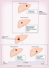
<b>Figure 1.</b>
Figure 1.. Schematic representation of two modalities of recurrences after local treatment of hepatocellular carcinoma.
Local relapse, defined as tumor emergence directly at the margin of the initial treatment zone, is linked either to: the stage and the biology of the tumor; the technical and technological conditions of ablation; factors related to underlying liver disease. Distant relapse, defined as separated by nontumor liver, is related either to metastases (usually before the 2-year cut-off time) or de novo carcinogenesis (usually after the 2-year cut-off time) and mainly linked to: staging and the biology of the tumor; and factors related to underlying liver disease. HCC: Hepatocellular carcinoma.
<b>Figure 2.</b>
Figure 2.. Diagrammatic representations of ablation areas induction with multiseparated applicators according the use of centrifugal (monopolar) or centripetal (multibipolar) energy deposition devices.
In this instance, for a tumor of roughly 3 cm in diameter, three applicators positioning are required with both devices to achieve complete ablation of the tumor and a minimal 5 mm free margin. On the left, with monopolar centrifugal energy diffusion devices (monopolar radiofrequency ablation (RFA), microwave, cryotherapy and laser), three overlapped single ablation are needed (A). Ablations can be performed sequentially with a single applicator or simultaneously with three applicators. On the right, with multibipolar centripetal energy diffusion devices (multibipolar RFA or electroporation) the same results could be more reliably achieved by single-bloc ablation implying the simultaneous use of three applicators. Each possible pair's combination of applicators is sequentially activated in bipolar mode. As shown in the diagram, below a maximal tumor diameter depending mainly on the technical characteristics of the devices used this type of multibipolar technology allows ‘no-touch’ ablation consisting to insert applicators outside of the tumor. Thus the achievement of free tumor margin becomes even more reliable because the maximum amount of energy is actively deposited in the margin conversely to monopolar devices which induce ablation of the margin at the end stage of the procedure by passive rapidly decreasing energy diffusion from the center to the periphery. (B–E): Case of typical complete ablation of small hepatocellular carcinoma (HCC; 3 cm) with proper free margin (1–2 cm) achieved with single no-touch multibipolar RFA. Before treatment, CT shows typical HCC (arrows) with at arterial phase (B, axial view) and washout at portal phase (C, frontal view). One month after treatment there is no more hyperenhancing tumor tissue at arterial phase (D, axial view) but the ‘ghost’ image of the nodule (arrows) within a large ablation area including a 1 cm minimal free margin even shows at portal phase in orthogonal plan (E, frontal view).
<b>Figure 3.</b>
Figure 3.. Touch imprints of hepatocellular carcinoma biospy samples.
One tumor biopsy sample is fixed in formalin (1), paraffin embedded (2) and sections are used for histological and immunohistochemical analysis (3). A second tumor biopsy sample (5) is apposed on sterile glass slides (4) before being snap frozen in liquid nitrogen (5). After staining, cytological analysis of the touched imprints (4) allows to affirm the presence of tumor in the frozen sample.
<b>Figure 4.</b>
Figure 4.. ESM-1 or Endocan immunostaining in hepatocellular carcinoma.
(A) Several cells lining tumor hepatocytes showed a strong cytoplasmic staining (arrows). (B) At a higher magnification, the elongated shape of the cells along with their sinusoidal localization and small fusiform nuclei allow identifying positive cells as tumor stromal endothelial cells. Tumor cells were negative. (C & D) In our experience, no staining at all was observed in 60% of early-stage uninodular hepatocellular carcinoma.

References

    1. Bruix J, Sherman M. Management of hepatocellular carcinoma: an update. Hepatology. 2011;53:1020–1022. - PMC - PubMed
    1. Lencioni RA, Allgaier HP, Cioni D, et al. Small hepatocellular carcinoma in cirrhosis: randomized comparison of radio-frequency thermal ablation versus percutaneous ethanol injection. Radiology. 2003;228:235–240. - PubMed
    1. Lin SM, Lin CJ, Lin CC, Hsu CW, Chen YC. Randomised controlled trial comparing percutaneous radiofrequency thermal ablation, percutaneous ethanol injection, and percutaneous acetic acid injection to treat hepatocellular carcinoma of 3 cm or less. Gut. 2005;54:1151–1156. - PMC - PubMed
    1. Shiina S, Teratani T, Obi S, et al. A randomized controlled trial of radiofrequency ablation with ethanol injection for small hepatocellular carcinoma. Gastroenterology. 2005;129:122–130. - PubMed
    1. Tiong L, Maddern GJ. Systematic review and meta-analysis of survival and disease recurrence after radiofrequency ablation for hepatocellular carcinoma. Br. J. Surg. 2011;98:1210–1224. - PubMed

LinkOut - more resources