. 2018 Sep 7;13(9):e0203426.
doi: 10.1371/journal.pone.0203426. eCollection 2018.

Adipose-derived stem cell-mediated paclitaxel delivery inhibits breast cancer growth

Affiliations

Adipose-derived stem cell-mediated paclitaxel delivery inhibits breast cancer growth

Maria Giovanna Scioli et al. PLoS One. .

Abstract

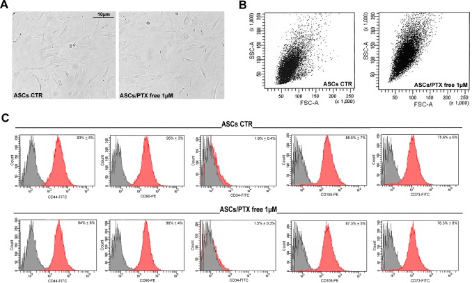
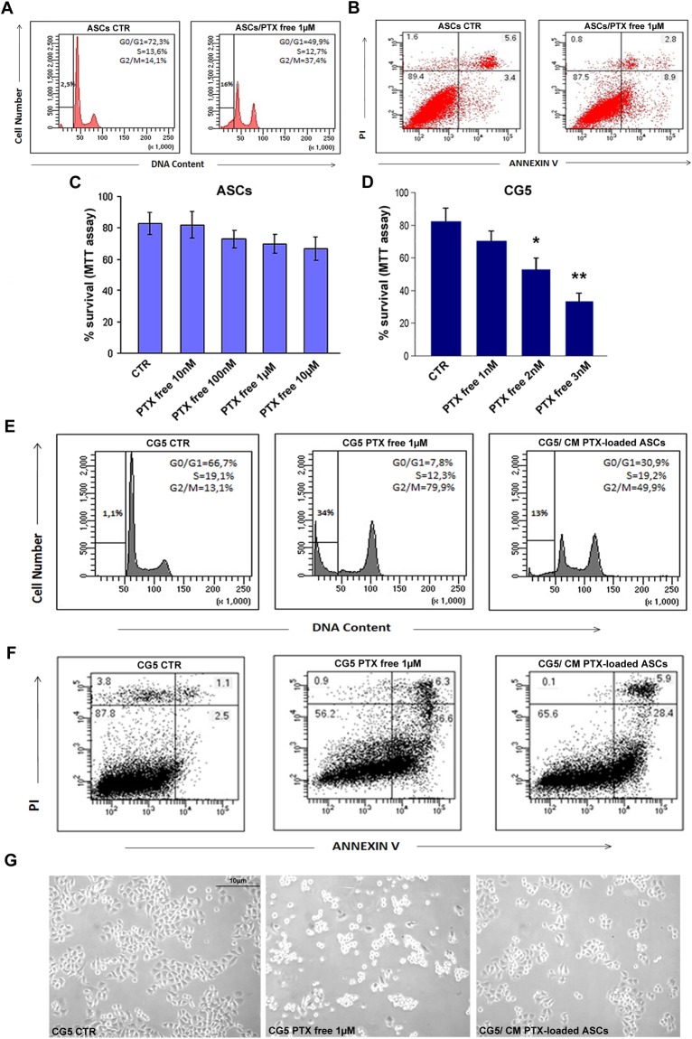
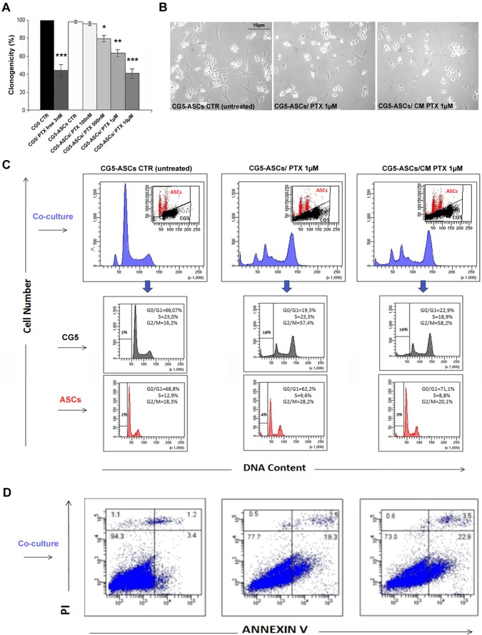
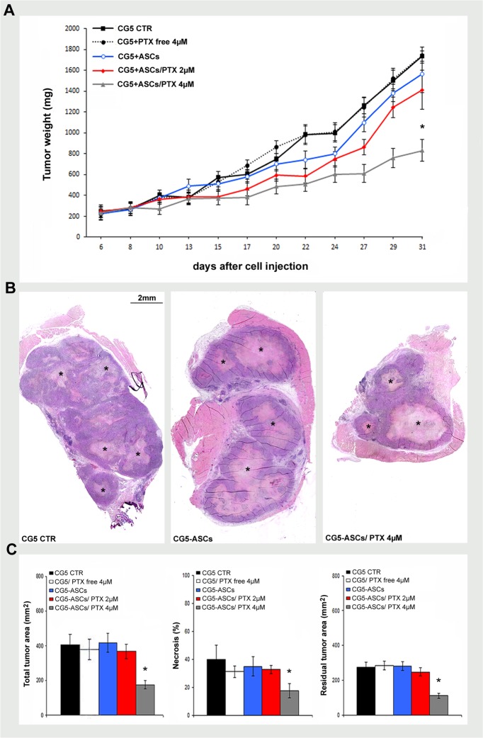
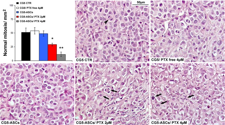
Breast cancer represents the main malignancy in women and autologous fat grafting is a diffuse procedure in the management of post-surgical breast defects causing patients' psychosocial problems, with high costs for the public health. Recently, beneficial effects of fat grafting during post-surgical breast reconstruction have been amplified from the enrichment with human adipose-derived stem cells (ASCs) present in the stromal vascular fraction (SVF) of adult adipose tissue isolated during intraoperatory procedures. The major concern about the ASC enrichment during post-surgery breast reconstruction depends on their potential ability to release growth factors and hormones that can promote proliferation of residual or quiescent cancer cells, with the risk of de novo cancer development or recurrence. The recent description that adult stem cells primed in vitro may be vehicle for anti-cancer drug delivery offers a new vision concerning the role of ASCs in breast reconstruction after cancer surgery. Paclitaxel (PTX) is a chemotherapeutic agent acting as a microtubule-stabilizing drug inhibiting cancer cell mitotic activity. We optimized PTX loading and release in cultured ASCs and then analyzed the effects of PTX-loaded ASCs and their conditioned medium on CG5 breast cancer survival, proliferation and apoptosis in vitro, and inCG5 xenograft in vivo. We documented that ASCs can uptake and release PTX in vitro, with slight cytotoxic effects. Interestingly, PTX-loaded ASCs in co-culture, as well as conditioned medium alone, inhibited CG5 cell proliferation and survival in vitro and xenograft tumor growth in vivo. The antitumor effect of PTX-loaded ASCs may offer a new perspective concerning the use of ASCs during breast reconstruction becoming an additional local preventive chemotherapeutic agent against tumor recurrence. However, further experiments in vitro and in vivo are needed to collect more evidence confirming the efficacy and safety in cancer patients.


Conflict of interest statement

The authors have declared that no competing interests exist.

Figures

Fig 1
Fig 1. Morphology and stem immunophenotype of PTX treated ASCs.
A) Representative images of cultured ASCs treated with PTX 1μM for 6 hours. B) Flow cytometric analysis of side and forward scatter of control (CTR) and PTX-loaded ASCs (ASCs/PTX; 1μM for 6 hours) showing an increased intracytoplasmic granularity of PTX-loaded ASCs. C) Histograms showing similar percentages of CD90, CD44, CD34, CD105 and CD73 positive cells in CTR (untreated) and PTX treated ASCs. The grey histograms indicate isotype controls, while the red histograms antibody expression.
Fig 2
Fig 2. Cell cycle, survival and apoptosis of PTX treated ASCs and CG5.
A) Cell cycle analysis by flow cytometry of CTR (untreated) and PTX treated ASCs (1μM for 6 hours). Adherent and not adherent cells were processed for PI staining to evaluate cell cycle. B) Cytofluorimetric bi-parametric analysis of the annexin V versus PI staining assay. The percentage reported in the annexin V+/PI+ region of each histogram represents the apoptotic cells. C, D) MTT assay of ASCs and CG5 treated with different concentrations of PTX for 6 hours, respectively. E) CG5 cells were treated with PTX free at 1 μM or with conditioned medium previously obtained by PTX primed ASCs at 1 μM. Adherent and not adherent cells were processed for cell cycle analysis. The percentages of cells in the different phases of cell cycle were reported inside the relative histograms. F) Cytofluorimetric bi-parametric analysis of the annexin V versus PI staining assay. G) Representative images of CG5 cell cultures with different treatments. Abbreviation: CM, conditioned medium.t-test: * and ** indicate p< 0.01 and p< 0.001, respectively.
Fig 3
Fig 3. Clonogenicity, cell cycle and apoptosis of CG5/ASC co-cultures in the presence of PTX.
A) The inhibitory activity of PTX-loaded ASCs on CG5 proliferation by clonogenic assay. ASCs were primed or not with different concentrations of PTX (for 6 hours), mixed with CG5 and the number of colonies evaluated compared with CG5 alone or treated with PTX free (3nM). B) Representative images of CG5 cell cultures with different treatments. C) Cell cycle analysis by flow cytometry of CG5/ASC co-cultures with different treatments (untreated ASCs, PTX-loaded ASCs or CM PTX-loaded ASCs at 1μM). The two cell populations were separated by analytical sorter in bi-parametric analysis of DNA content versus forward scatter. D) Cytofluorimetric bi-parametric analysis of the annexin V versus PI staining assay. The percentage reported in the annexin V+/PI+ region of each histogram represents the apoptotic cells. Abbreviation: CM, conditioned medium.t-test: *, ** and *** indicate p< 0.05, p< 0.01 and p< 0.001, respectively.
Fig 4
Fig 4. The therapeutic efficacy of PTX-primed ASCs on tumor growth in vivo.
A) Tumor growth curve of CG5 cells (1x10⁶) co-injected i.m.in nude mice (5 mice/group) with ASCs (1x10⁶) unloaded or primed with PTX (2μM or 4μM for 6 hours), ratio of 1:1. Other control groups were CG5 alone or mixed with PTX free (4μM). B, C) Representative histological images and morphometric analysis of tumor xenografts showing total tumor area, percentage of necrosis, residual tumor area (mm2). Necrotic area, light pink eosinophilic area inside tumor mass (asterisk). t-test: *, p< 0.05.
Fig 5
Fig 5. The effects of PTX-primed ASCs on tumor mitotic activity in vivo.
Mitotic activity in high power field images of CG5 xenograft showing normal (bipolar, arrowhead) and aberrant mitosis (monoastral spindles and multipolar spindles, arrows). t-test: * and **, p< 0.05 and p< 0.01, respectively.

References

    1. Coleman SR, Saboeiro AP. Fat grafting to the breast revisited: safety and efficacy. Plast Reconstr Surg. 2007; 119:775–785. 10.1097/01.prs.0000252001.59162.c9 - DOI - PubMed
    1. Haller H, Winkler MM, Klose P, Dobos G, Kümmel S, Cramer H. Mindfulness-based interventions for women with breast cancer: an updated systematic review and meta-analysis. Acta Oncol. 2017;56:1665–1676. 10.1080/0284186X.2017.1342862 - DOI - PubMed
    1. Gentile P, Orlandi A, Scioli MG, Di Pasquali C, Bocchini I, Cervelli V. Concise review: adipose-derived stromal vascular fraction cells and platelet-rich plasma: basic and clinical implications for tissue engineering therapies in regenerative surgery. Stem Cells Transl Med. 2012; 1:230–236. 10.5966/sctm.2011-0054 - DOI - PMC - PubMed
    1. Zuk PA, Zhu M, Mizuno H, Huang J, Futrell JW, Katz AJ, et al. Multilineage cells from human adipose tissue: implications for cell-based therapies. Tissue Eng. 2001; 7:211–228. 10.1089/107632701300062859 - DOI - PubMed
    1. Gentile P, Orlandi A, Scioli MG, Di Pasquali C, Bocchini I, Curcio CB, et al. A comparative translational study: the combined use of enhanced stromal vascular fraction and platelet-rich plasma improves fat grafting maintenance in breast reconstruction. Stem Cells Transl Med. 2012; 1:341–351. 10.5966/sctm.2011-0065 - DOI - PMC - PubMed

MeSH terms

Substances