. 2018 Sep 6;71(5):745-760.e5.
doi: 10.1016/j.molcel.2018.07.034.

Non-canonical Activation of the DNA Sensing Adaptor STING by ATM and IFI16 Mediates NF-κB Signaling after Nuclear DNA Damage

Affiliations

Non-canonical Activation of the DNA Sensing Adaptor STING by ATM and IFI16 Mediates NF-κB Signaling after Nuclear DNA Damage

Gillian Dunphy et al. Mol Cell. .

Abstract

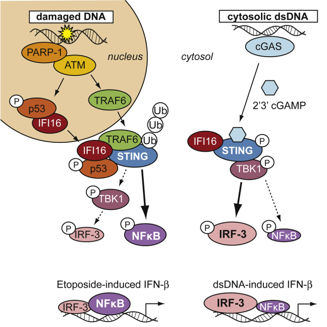
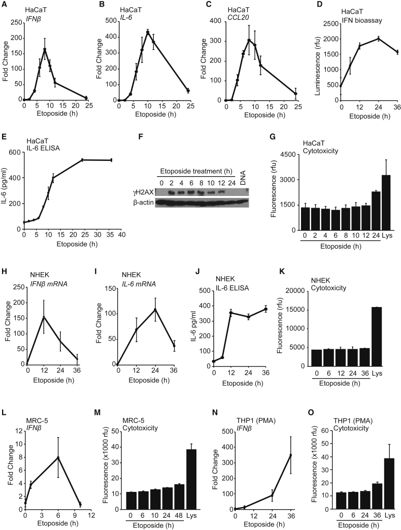
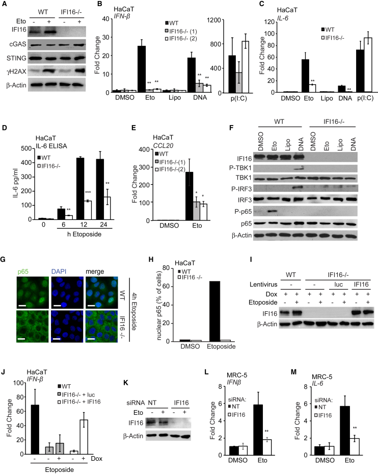
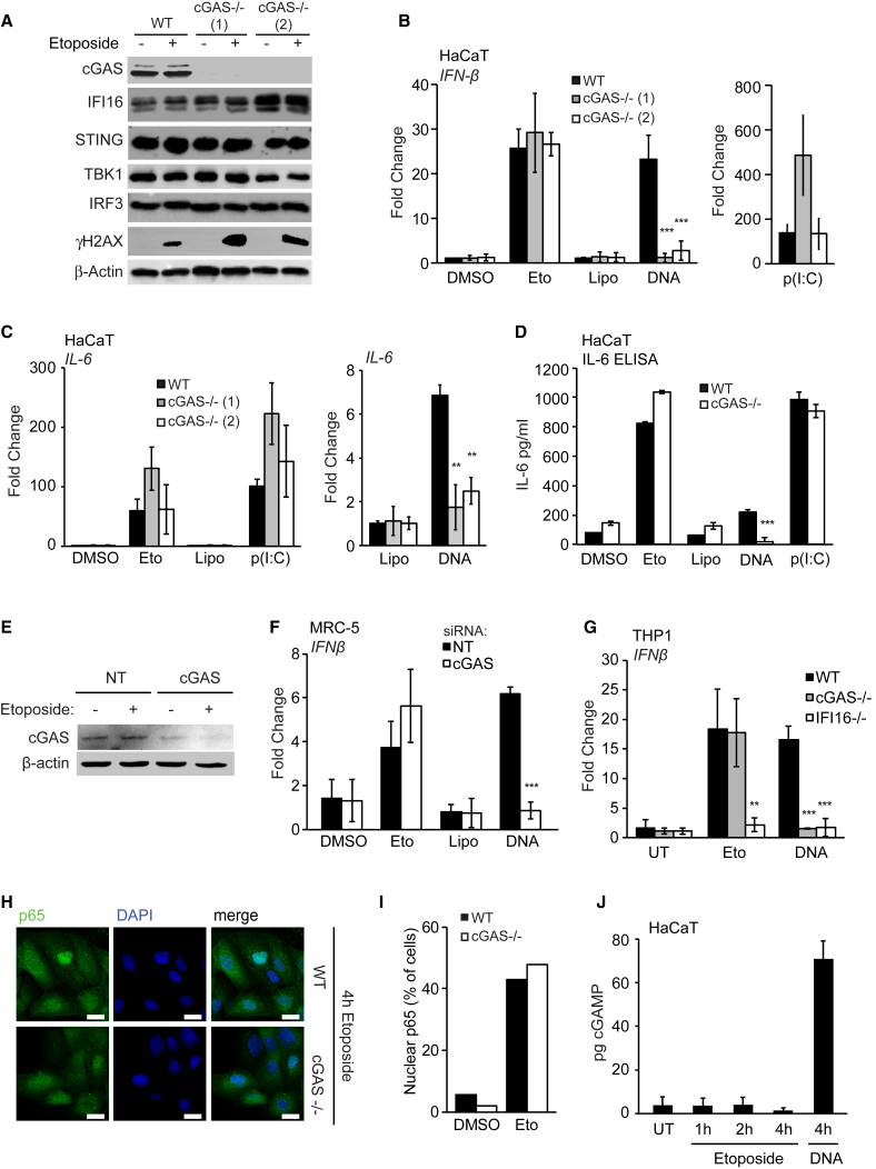
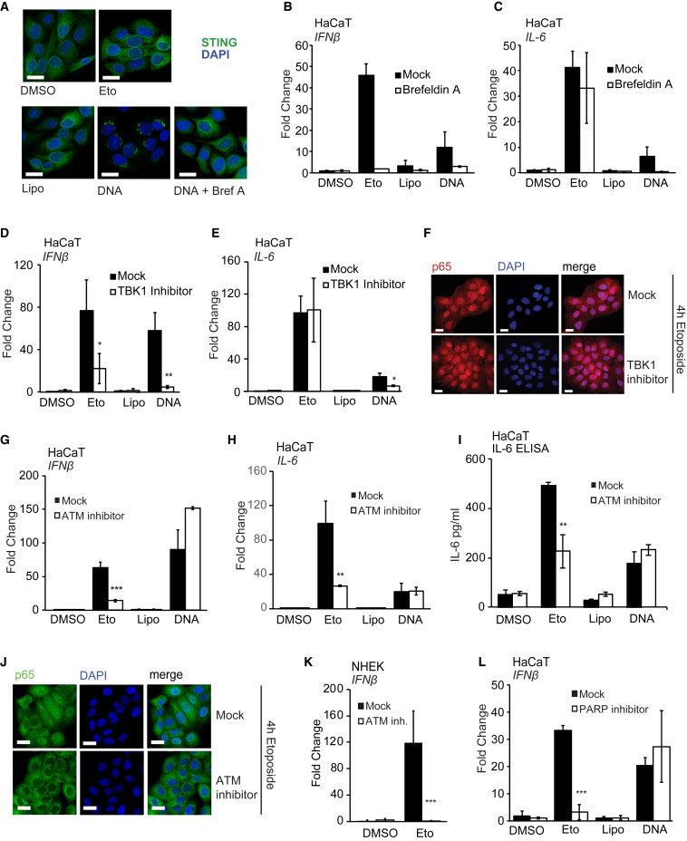
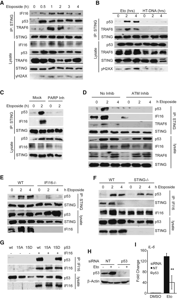
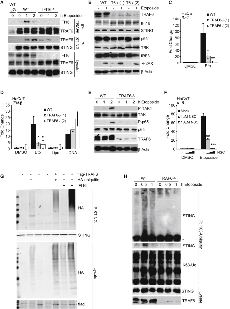
DNA damage can be sensed as a danger-associated molecular pattern by the innate immune system. Here we find that keratinocytes and other human cells mount an innate immune response within hours of etoposide-induced DNA damage, which involves the DNA sensing adaptor STING but is independent of the cytosolic DNA receptor cGAS. This non-canonical activation of STING is mediated by the DNA binding protein IFI16, together with the DNA damage response factors ATM and PARP-1, resulting in the assembly of an alternative STING signaling complex that includes the tumor suppressor p53 and the E3 ubiquitin ligase TRAF6. TRAF6 catalyzes the formation of K63-linked ubiquitin chains on STING, leading to the activation of the transcription factor NF-κB and the induction of an alternative STING-dependent gene expression program. We propose that STING acts as a signaling hub that coordinates a transcriptional response depending on its mode of activation.

Keywords: DNA damage; IFI16; STING; TRAF6; etoposide; innate immunity; interferon; p53; ubiquitin.


Figures

None
Graphical abstract
Figure 1
Figure 1
Etoposide-Mediated DNA Damage Induces an Acute Innate Immune Response in Human Cells (A–C) HaCaT keratinocytes were treated with 50 μM etoposide for the times indicated before qRT-PCR analysis of IFN-β (A), IL-6 (B), and CCL20 (C) mRNA. (D and E) Supernatants from cells treated with 50 μM etoposide were analyzed for secreted type I IFN using a bio-assay (D) or IL-6 protein using ELISA (E). (F) HaCaT cells were treated with 50 μM etoposide for the times indicated or transfected with 1 μg/mL herring testis (HT)-DNA for 6 hr. Phosphorylation of γH2A.X was analyzed by immunoblotting. (G) Cytotoxicity assay of HaCaT cells treated with 50 μM etoposide for the times indicated or lysed (Lys). (H and I) Primary normal human epidermal keratinocytes (NHEKs) from adult donors were treated with 50 μM etoposide for the times indicated before qRT-PCR analysis of IFN-β (H) and IL-6 (I) mRNA. (J) Supernatants from NHEK cells treated as in (H) were analyzed for IL-6 secretion by ELISA. (K) Cytotoxicity assay of NHEK cells treated as in (H) or lysed (Lys). (L) Primary MRC-5 fibroblasts were treated with 50 μM etoposide before qRT-PCR analysis of IFN-β mRNA expression. (M) Cytotoxicity assay of MRC-5 cells treated with 50 μM etoposide or lysed (Lys). (N) PMA-differentiated THP1 cells were stimulated with 50 μM etoposide for indicated times before qRT-PCR analysis of IFN-β mRNA. (O) Cytotoxicity assay of THP1 cells treated as in (N) or lysed (Lys). Data are presented as mean values of biological triplicates ± SD. See also Figure S1.
Figure 2
Figure 2
STING Is Required for the Innate Immune Response to Etoposide-Induced DNA Damage (A) Wild-type (WT) and STING−/− HaCaT cells were treated with DMSO or 50 μM etoposide for 6 hr, and protein expression was analyzed by immunoblotting. (B) Clonogenic survival assay of WT and STING−/− HaCaT cells. Numbers of colonies > 50 cells were counted and expressed as a percentage of untreated control. (C and D) WT HaCaT and two STING−/− clones were treated with DMSO or 50 μM etoposide, mock transfected (Lipo), or transfected with 1 μg/mL HT-DNA or 100 ng/mL poly(I:C) for 6 hr before qRT-PCR analysis of IFN-β (C) and IL-6 (D) mRNA expression. (E) ELISA analysis of IL-6 secretion in supernatants from cells treated as in (C) for 24 hr. (F) qRT-PCR array analysis of cytokine and chemokine expression in WT and STING−/− HaCaT cells treated with DMSO, 50 μM etoposide, Lipofectamine, or 1 μg/mL HT-DNA for 6 hr. Shown are genes induced at least 2-fold over controls. (G and H) WT and STING−/− HaCaT cells grown on coverslips were treated with 50 μM etoposide for 4 hr and stained for NF-κB p65 (green) and DNA (DAPI, blue) for analysis by confocal microscopy (G) and quantification of p65 nuclear translocation (H). Scale bar, 20 μm. (I and J) NHEKs were treated with non-targeting (NT) or STING-targeting siRNA pools for 48 hr before treatment with 50 μM etoposide for 24 hr. STING protein levels were analyzed by immunoblotting (I), and IFN-β mRNA expression was quantified by qRT-PCR (J). (K) MRC-5 fibroblasts were treated with non-targeting (NT) or STING-targeting siRNA pools for 48 hr before treatment with 50 μM etoposide for 6 hr and analysis of IFN-β mRNA by RT-PCR. (L) PMA-differentiated WT and STING−/− THP1 cells were stimulated with 50 μM etoposide for 30 hr or 1 μg/mL HT-DNA for 6 hr before qRT-PCR analysis of IFN-β mRNA. Data are presented as mean values of biological triplicates ± SD. See also Figures S2 and S3A–S3F.
Figure 3
Figure 3
The Innate Immune Response to Etoposide-Induced Damage Involves IFI16 (A) Immunoblotting analysis of WT and IFI16−/− HaCaT cells stimulated with 50 μM etoposide or DMSO for 6 hr. (B and C) WT HaCaT cells and two IFI16−/− cell clones were treated for 6 hr with DMSO or 50 μM etoposide, mock transfected (Lipo), or transfected with 1 μg/mL HT-DNA or 100 ng/mL poly(I:C). IFN-β (B) or IL-6 (C) mRNA was quantified by qRT-PCR. (D) ELISA analysis of IL-6 protein in supernatants from WT and IFI16−/− HaCaT cells treated with 50 μM etoposide for indicated times. (E) qRT-PCR analysis of CCL20 mRNA in WT and IFI16−/− HaCaT cells treated with DMSO or 50 μM etoposide for 6 hr. (F) WT and IFI16−/− HaCaT cells were treated as in (B) for 4 hr before analysis of protein expression by immunoblotting. (G) WT and IFI16−/− HaCaT cells grown on coverslips were treated with 50 μM etoposide for 4 hr and fixed and stained for p65 (green) and DNA (DAPI, blue). Scale bar, 20 μm. (H) Quantification of p65 nuclear translocation in cells from (G). (I) Immunoblotting analysis of WT HaCaT cells and IFI16−/− HaCaT cells reconstituted with lentiviruses for the expression of Luciferase (luc) or IFI16 as indicated. Cells were treated with doxycycline for 24 hr to induce expression and then stimulated with 50 μM etoposide for 6 hr. (J) qRT-PCR analysis of IFN-β mRNA in cells treated as in (I) as indicated. (K–M) MRC-5 fibroblasts treated with non-targeting (NT) or IFI16-targeting siRNA pools for 48 hr before treatment with 50 μM etoposide or DMSO for 6 hr. IFI16 protein expression was analyzed by immunoblotting (K). IFN-β (L) and IL-6 (M) mRNA levels were analyzed by qRT-PCR. Data are presented as mean values of biological triplicates ± SD. See also Figures S3G–S3L.
Figure 4
Figure 4
cGAS Is Dispensable for the Early Innate Immune Response to Nuclear DNA Damage (A) Immunoblotting analysis of WT and two cGAS−/− HaCaT clones treated with DMSO or 50 μM etoposide for 6 hr. (B and C) WT and cGAS−/− HaCaT cells were treated with DMSO or 50 μM etoposide, mock transfected (Lipo), or transfected with 1 μg/mL HT-DNA or 100 ng/mL poly(I:C) for 6 hr before qRT-PCR analysis of IFN-β (B) and IL-6 (C) mRNA expression. (D) IL-6 in supernatants from WT and cGAS−/− HaCaT cells treated with 50 μM etoposide quantified by ELISA. (E) MRC-5 fibroblasts were treated with non-targeting (NT) or cGAS-targeting siRNA pools for 48 hr before treatment with 50 μM etoposide for 6 hr. cGAS protein expression was analyzed by western blot. (F) qRT-PCR analysis of IFN-β mRNA expression in MRC-5 fibroblasts treated with siRNA as in (E) and stimulated with 50 μM etoposide or transfected with 1 μg/mL HT-DNA for 6 hr. (G) PMA-differentiated WT, cGAS−/−, and IFI16−/− THP1 cells were treated with 50 μM etoposide for 30 hr or 1 μg/mL HT-DNA for 6 hr before qRT-PCR analysis of IFN-β mRNA. (H) WT and cGAS−/− HaCaT cells grown on coverslips were treated with 50 μM etoposide for 4 hr, stained for p65 (green) and DNA (DAPI, blue), and visualized by confocal microscopy. Scale bar, 20 μm. (I) Quantification of p65 translocation from (H). (J) HaCaT cells were treated with 50 μM etoposide for the indicated times or transfected with 1 μg/mL HT-DNA for 4 hr. cGAMP production was quantified by LC-MS. Data are presented as mean values of biological triplicates ± SD. See also Figure S4.
Figure 5
Figure 5
Etoposide-Induced NF-κB Activation Involves DNA Damage Factors, but Not TBK1 Activity (A) HaCaT cells grown on coverslips were pre-treated for 30 min with 3 μg/mL brefeldin A where indicated before stimulation with 50 μM etoposide or transfection of 1 μg/mL HT-DNA. Cells were fixed and stained for STING (green) and DNA (DAPI, blue). Scale bar, 20 μm. (B and C) HaCaT cells were pre-treated for 30 min with 3 μg/mL brefeldin A before treatment with 50 μM etoposide or DMSO, mock transfection (Lipo), or transfection of 1 μg/mL HT-DNA for 6 hr. IFN-β (B) and IL-6 (C) mRNA expression was analyzed by qRT-PCR. (D and E) HaCaT cells were pre-treated for 1 hr with 2 μM TBK1 inhibitor MRT67307 and stimulated as in (B) before qRT-PCR analysis of IFN-β (D) and IL-6 (E) mRNA expression. (F) HaCaT cells grown on coverslips were pre-treated with 2 μM TBK1 inhibitor MRT67307 for 1 hr before 4 hr of stimulation with 50 μM etoposide. Cells were fixed and stained for p65 (red) and DNA (DAPI, blue). Scale bar, 20 μm. (G and H) HaCaT cells were pre-treated for 1 hr with 10 μM ATM inhibitor KU55933 and stimulated as in (B). IFN-β (G) and IL-6 (H) mRNA expression was quantified by qRT-PCR. (I) ELISA analysis of IL-6 secretion in supernatants from cells treated as in (G) and stimulated for 24 hr. (J) HaCaT cells grown on coverslips were pre-treated for 1 hr with 10 μM KU55933 before 4 hr of stimulation with 50 μM etoposide. Cells were fixed and stained for p65 (green) and DNA (DAPI, blue). Scale bar, 20 μm. (K) qRT-PCR analysis of IFN-β mRNA expression in NHEK cells pre-treated for 1 hr with 10 μM KU55933, followed by treatment with 50 μM etoposide for 24 hr. (L) qRT-PCR analysis of IFN-β mRNA in HaCaT cells pre-treated for 1 hr with 10 μM PARP inhibitor PJ34 before treatment as in (B) for 6 hr. Data are presented as mean values of biological triplicates ± SD. See also Figure S5.
Figure 6
Figure 6
Nuclear DNA Damage Results in the Assembly of a Non-canonical Signaling Complex Containing STING (A) Immunoprecipitation of STING from HaCaT cells treated with 50 μM etoposide for the indicated times. Immunoprecipitates (IPs) and whole-cell lysates were analyzed by immunoblotting. (B) Immunoblotting analysis following immunoprecipitation of STING from HaCaT cells treated with 50 μM etoposide or transfected with 1 μg/mL HT-DNA as indicated. (C) Immunoprecipitation of STING from HaCaT cells pre-treated for 1 hr with 10 μM PARP inhibitor PJ34, followed by treatment with 50 μM etoposide for 2 hr. (D) Immunoprecipitation of STING from HaCaT cells pre-treated for 1 hr with 10 μM ATM inhibitor KU55933 followed by treatment with 50 μM etoposide. (E) Immunoprecipitation of STING from WT and IFI16−/− HaCaT cells treated with 50 μM etoposide as indicated. (F) Immunoprecipitation of IFI16 from WT and STING−/− HaCaT cells treated with 50 μM etoposide as indicated. (G) HEK293T cells transfected with expression constructs for IFI16 and WT p53 or the S15A or S15D p53 mutants as indicated. 24 hr after transfection, IFI16 was immunoprecipitated from lysates. (H) p53 protein levels in HaCaT cells transfected with a non-targeting (NT) or a p53-targeting siRNA pool for 48 hr before stimulation with 50 μM etoposide for 6 hr. (I) qRT-PCR analysis of IL-6 mRNA expression in cells treated as in (H). See also Figure S6.
Figure 7
Figure 7
TRAF6 Mediates the K63-Linked Poly-ubiquitylation of STING (A) Immunoprecipitation of TRAF6 and STING from WT and IFI16−/− HaCaT cells treated with 50 μM etoposide as indicated. Immunoprecipitates (IP) with immunoglobulin G (IgG) control and input lysates were analyzed by immunoblotting. (B) WT and two TRAF6−/− HaCaT clones were treated with 50 μM etoposide for 6 hr, and protein expression was analyzed by immunoblotting. (C) qRT-PCR analysis of IL-6 mRNA expression in cells treated as in (B). (D) WT and TRAF6−/− HaCaT cells were treated with 50 μM etoposide or DMSO, mock transfected (Lipo), or transfected with 1 μg/mL HT-DNA for 6 hr before qRT-PCR analysis of IFN-β mRNA. (E) Immunoblotting analysis of WT and TRAF6−/− HaCaT cells treated with 50 μM etoposide for the indicated times. (F) HaCaT cells were pre-treated for 1 hr with the indicated concentrations of Ubc13 inhibitor NSC697923 (NSC) before 6 hr of stimulation with 50 μM etoposide. IL-6 mRNA expression was quantified by qRT-PCR. (G) HEK293T cells were transfected with plasmids for the expression of IFI16, FLAG-tagged TRAF6, and hemagglutinin (HA)-tagged ubiquitin as indicated. 24 hr after transfection, STING was immunoprecipitated, and proteins in immunoprecipitates and input lysates were analyzed by immunoblotting. (H) Immunoprecipitation of K63-linked ubiquitin chains from WT and TRAF6−/− HaCaT cells treated with 50 μM etoposide for the times indicated. Higher molecular weight forms of modified STING are visualized by gradient SDS-PAGE above the antibody heavy chain (), top panel, together with the association of unmodified STING, lower panel. See also Figure S7.

References

    1. Abe T., Barber G.N. Cytosolic-DNA-mediated, STING-dependent proinflammatory gene induction necessitates canonical NF-κB activation through TBK1. J. Virol. 2014;88:5328–5341. - PMC - PubMed
    1. Aglipay J.A., Lee S.W., Okada S., Fujiuchi N., Ohtsuka T., Kwak J.C., Wang Y., Johnstone R.W., Deng C., Qin J., Ouchi T. A member of the Pyrin family, IFI16, is a novel BRCA1-associated protein involved in the p53-mediated apoptosis pathway. Oncogene. 2003;22:8931–8938. - PubMed
    1. Almine J.F., O’Hare C.A.J., Dunphy G., Haga I.R., Naik R.J., Atrih A., Connolly D.J., Taylor J., Kelsall I.R., Bowie A.G. IFI16 and cGAS cooperate in the activation of STING during DNA sensing in human keratinocytes. Nat. Commun. 2017;8:14392. - PMC - PubMed
    1. Banin S., Moyal L., Shieh S., Taya Y., Anderson C.W., Chessa L., Smorodinsky N.I., Prives C., Reiss Y., Shiloh Y., Ziv Y. Enhanced phosphorylation of p53 by ATM in response to DNA damage. Science. 1998;281:1674–1677. - PubMed
    1. Boukamp P., Petrussevska R.T., Breitkreutz D., Hornung J., Markham A., Fusenig N.E. Normal keratinization in a spontaneously immortalized aneuploid human keratinocyte cell line. J. Cell Biol. 1988;106:761–771. - PMC - PubMed

Publication types

MeSH terms