

Real-time non-invasive in vivo visible light detection of cortical spreading depolarizations in mice
- PMID: 30194041
- PMCID: PMC6214472
- DOI: 10.1016/j.jneumeth.2018.09.001
Real-time non-invasive in vivo visible light detection of cortical spreading depolarizations in mice
Abstract
Background: Cortical spreading depolarization (CSD) is a phenomenon classically associated with migraine aura. CSDs have also been implicated in secondary injury following ischemic stroke, intracerebral hemorrhage, subarachnoid hemorrhage, and traumatic brain injury; however, most investigations involving these disease processes do not account for the occurrence of CSDs. A major barrier to detection of CSDs in experimental models is that currently validated methods are invasive and require specialized equipment and a high level of expertise to implement.
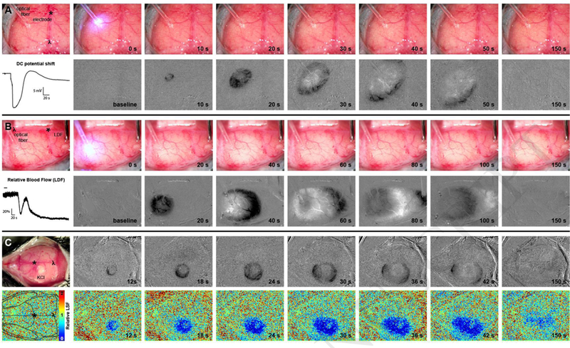
New method: We present a low-cost, easy-to-implement approach to the detection of CSDs in the mouse through full-thickness intact skull. Our method uses the optical intrinsic signal from white light illumination (OIS-WL) and allows for real-time in vivo detection of CSDs using readily available USB cameras.
Results: OIS-WL detected 100% of CSDs that were seen with simultaneous electrode recording (69 CSDs in 28 mice), laser Doppler flowmetry (82 CSDs in 10 mice), laser speckle flowmetry (68 CSDs in 25 mice), or combined electrode recording plus laser speckle flowmetry (29 CSDs in 20 mice). OIS-WL detected 1 additional CSD that was missed by laser Doppler flowmetry.
Comparison with existing methods: OIS-WL is less invasive than electrophysiological recordings and easier to implement than laser speckle flowmetry. Moreover, it provides excellent spatial and temporal resolution for dynamic imaging of CSDs in the setting of brain injury.
Conclusions: Detection of CSDs with an inexpensive USB camera and white light source provides a reliable method for the in vivo and non-invasive detection of CSDs through unaltered mouse skull.
Keywords: Cortical spreading depression; Migraine; Non-invasive; Optical intrinsic signal imaging; Stroke.
Copyright © 2018 Elsevier B.V. All rights reserved.
Figures
References
-
- Ayata C (2013) Pearls and pitfalls in experimental models of spreading depression. Cephalalgia : an international journal of headache 33:604–613. - PubMed
-
- Dunn AK, Bolay H, Moskowitz MA, Boas DA (2001) Dynamic imaging of cerebral blood flow using laser speckle. Journal of cerebral blood flow and metabolism : official journal of the International Society of Cerebral Blood Flow and Metabolism 21:195–201. - PubMed
Publication types
MeSH terms
Grants and funding
LinkOut - more resources
Full Text Sources
Other Literature Sources
Miscellaneous
