. 2018 Oct 1;215(10):2497-2508.
doi: 10.1084/jem.20180008. Epub 2018 Sep 7.

Endothelial cell fitness dictates the source of regenerating liver vasculature

Affiliations

Endothelial cell fitness dictates the source of regenerating liver vasculature

Mahak Singhal et al. J Exp Med. .

Abstract

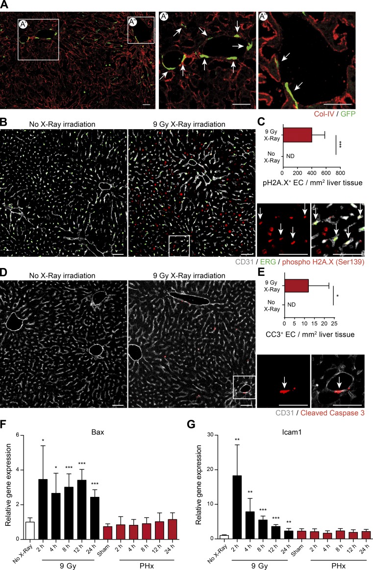
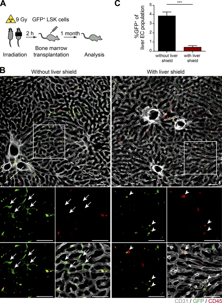
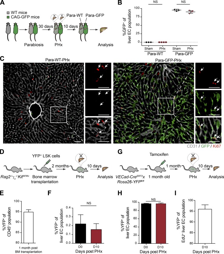
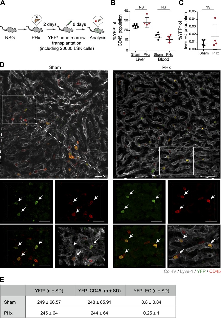
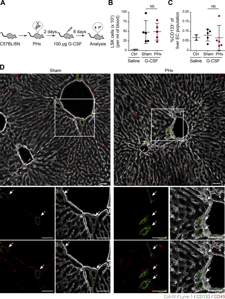
Neoangiogenesis plays a key role in diverse pathophysiological conditions, including liver regeneration. Yet, the source of new endothelial cells (ECs) remains elusive. By analyzing the regeneration of the liver vasculature in irradiation-based myeloablative and nonmyeloablative bone marrow transplantation mouse models, we discovered that neoangiogenesis in livers with intact endothelium was solely mediated by proliferation of resident ECs. However, following irradiation-induced EC damage, bone marrow-derived mononuclear cells were recruited and incorporated into the vasculature. Further experiments with direct bone marrow infusion or granulocyte colony-stimulating factor (G-CSF)-mediated progenitor cell mobilization, which resembles clinically relevant stem cell therapy, demonstrated that bone marrow-derived cells did not contribute to the regeneration of liver vasculature after two-thirds partial hepatectomy (PHx). Taken together, the data reconcile many of the discrepancies in the literature and highlight that the cellular source of regenerating endothelium depends on the fitness of the residual vasculature.


Figures

None
Graphical abstract
Figure 1.
Figure 1.
Irradiation-based myeloablation induces EC injury and primes for BMDMC incorporation. (A) Representative images of liver sections of irradiation-conditioned GFP+ bone marrow–transplanted sham-operated mice. (A’ and A’’) Zoomed-in images illustrating GFP+ cells incorporated into the liver vasculature. Arrows indicate GFP+ ECs. Scale bars, 100 µm. For complete confocal reconstruction, see Video 1. (B) Representative images of liver sections of control or irradiated mice costained with phospho-H2A.X (Ser139), CD31 (EC-specific surface marker), and ERG (EC-specific nuclear marker). Zoomed-in images are shown on the right. Arrows indicate phospho-H2A.X (Ser139)–positive ECs. Scale bars, 50 µm. (C) The plot shows the count of pH2A.X+ ECs per 1 mm2 of liver tissue (mean ± SD, n = 6 mice). (D) Representative images of liver sections of control or irradiated mice costained with cleaved caspase-3 (CC3) and CD31 (EC-specific surface marker). Zoomed-in images are shown on the right. Arrows indicate CC3+ ECs. Scale bars, 50 µm. (E) The plot shows the count of CC3+ ECs per 1 mm2 of liver tissue (mean ± SD, n = 6 mice). (F and G) Quantitative PCR analysis of mRNA expression of Bax (F) and Icam1 (G) in livers of mice after irradiation or PHx (mean ± SD, n = 5–6 mice for each time point). ND, nondetectable; *, P < 0.05; **, P < 0.01; ***, P < 0.001 (two-tailed Student’s t test).
Figure 2.
Figure 2.
A radioprotective shield reduces the recruitment of BMDMCs to the liver vasculature. (A) Experimental outline of PHx-induced liver regeneration in irradiation-conditioned GFP+ bone marrow–transplanted mice in the absence or presence of a liver shield. (B) Mice were irradiated with or without liver shield. Representative images of liver sections of bone marrow chimeric mice costained with GFP, CD45, and CD31 (EC-specific surface marker). Zoomed-in images are shown at the bottom. Arrows indicate GFP+ ECs. Arrowheads indicate GFP+ hematopoietic cells in the shielded liver. Scale bars, 50 µm. (C) The percentage of GFP+ ECs in livers of mice irradiated in the presence or absence of a liver shield was analyzed by FACS (mean ± SD, n = 6 mice). ***, P < 0.001 (two-tailed Student’s t test).
Figure 3.
Figure 3.
BMDMCs do not contribute to the regeneration of liver vasculature in nonmyeloablative models. (A) Experimental outline of the parabiotic model. The circulatory systems of WT and CAG-GFP mice were surgically conjoined, and both mice were subjected to PHx to induce liver regeneration. (B) The ratio of GFP+ ECs in the livers of Para-WT (sham operated and PHx) as well as Para-GFP (sham operated and PHx) mice was analyzed by FACS (mean ± SD, n = 3–4 mice). (C) Representative images of liver sections of Para-WT-PHx or Para-GFP-PHx mice costained with Ki67, GFP, and CD31 (EC-specific surface marker). Zoomed-in images are shown on the right. Arrows indicate Ki67+ ECs. Scale bars, 50 µm. (D) Experimental outline for transplantation of LSK cells into Rag2−/−γc−/−KitW/Wv mice. (E) FACS analysis of donor chimerism in CD45+ cells from peripheral blood of Rag2−/−γc−/−KitW/Wv recipients (mean ± SD, n = 4 mice). (F) The percentage of YFP+ ECs in the livers of Rag2−/−γc−/−KitW/Wv mice before and 10 d after PHx was analyzed by FACS (mean ± SD, n = 4 mice). (G) Experimental outline of the VECad-CreERT2xRosa26-YFPfl/fl genetic labeling model. (H) The frequency of YFP+ ECs in livers of the same VECad-CreERT2xRosa26-YFPfl/fl mouse before and 10 d after PHx was analyzed by FACS (mean ± SD, n = 6 mice). (I) The proportion of YFP+ cells among the total proliferated liver ECs (as labeled by EdU) after PHx (mean ± SD, n = 6 mice). Two-tailed Student’s t test.
Figure 4.
Figure 4.
Infused bone marrow cells do not incorporate into the regenerating liver vasculature. (A) Experimental outline of PHx-induced liver regeneration in NSG mice. Prior to the angiogenic phase (on day 2 after PHx), mice were infused intravenously with YFP-labeled bone marrow cells as a regenerative cellular therapy. (B) The frequency of YFP+ hematopoietic cells (CD45+) in the liver and the peripheral blood of sham-operated and PHx mice was analyzed by FACS (mean ± SD, n = 4–5 mice). (C) The ratio of YFP+ ECs in the livers of sham-operated and PHx mice was analyzed by FACS (mean ± SD, n = 4–5 mice). (D) Representative images of liver sections of sham-operated and PHx mice costained with YFP, CD45, and liver EC–specific marker (Lyve-1/Col-IV). Zoomed-in images are shown at the bottom. Arrows indicate YFP+ cells. All traced YFP+ cells were positive for CD45 but negative for EC markers. Scale bars, 25 µm. (E) Shown are the absolute numbers of YFP+ cells, YFP+ hematopoietic cells, and YFP+ ECs counted per 1 mm2 of the liver tissue of sham-operated and PHx mice (mean ± SD, n = 4–5 mice). Two-tailed Student’s t test.
Figure 5.
Figure 5.
G-CSF–mobilized bone marrow cells do not incorporate into the regenerating liver vasculature. (A) Experimental outline of PHx-induced liver regeneration in C57BL/6N mice. Prior to the angiogenic phase (on day 2 after PHx), mice were injected subcutaneously with 100 µg G-CSF (as a regenerative therapy) to mobilize bone marrow–derived progenitor cells. (B) The frequency of circulating LSK cells in the peripheral blood of sham-operated and PHx mice was analyzed by FACS (mean ± SD, n = 5 mice). (C) The ratio of CD133+ ECs in the livers of sham-operated and PHx mice was analyzed by FACS (mean ± SD, n = 5 mice). (B and C) Saline-injected mice served as controls. (D) Representative images of liver sections of sham-operated and PHx mice costained with CD133 (progenitor cell marker), CD45, and liver EC–specific marker (Lyve-1/Col-IV). Zoomed-in images are shown at the bottom. Arrows indicate CD133+ cells. All traced CD133+ cells were negative for CD45 and EC markers. Scale bars, 50 µm. Two-tailed Student’s t test.

Comment in

Similar articles

Cited by

References

    1. Abbuehl J.P., Tatarova Z., Held W., and Huelsken J.. 2017. Long-term engraftment of primary bone marrow stromal cells repairs niche damage and improves hematopoietic stem cell transplantation. Cell Stem Cell. 21:241–255.e6. 10.1016/j.stem.2017.07.004 - DOI - PubMed
    1. Almeida-Porada G., Zanjani E.D., and Porada C.D.. 2010. Bone marrow stem cells and liver regeneration. Exp. Hematol. 38:574–580. 10.1016/j.exphem.2010.04.007 - DOI - PMC - PubMed
    1. Aoki M., Yasutake M., and Murohara T.. 2004. Derivation of functional endothelial progenitor cells from human umbilical cord blood mononuclear cells isolated by a novel cell filtration device. Stem Cells. 22:994–1002. 10.1634/stemcells.22-6-994 - DOI - PubMed
    1. Asahara T., Murohara T., Sullivan A., Silver M., van der Zee R., Li T., Witzenbichler B., Schatteman G., and Isner J.M.. 1997. Isolation of putative progenitor endothelial cells for angiogenesis. Science. 275:964–967. 10.1126/science.275.5302.964 - DOI - PubMed
    1. Augustin H.G., and Koh G.Y.. 2017. Organotypic vasculature: From descriptive heterogeneity to functional pathophysiology. Science. 357:eaal2379 10.1126/science.aal2379 - DOI - PubMed

Publication types