. 2018 Sep 7;9(1):3655.
doi: 10.1038/s41467-018-06057-9.

Necroptosis mediates myofibre death in dystrophin-deficient mice

Affiliations

Necroptosis mediates myofibre death in dystrophin-deficient mice

Jennifer E Morgan et al. Nat Commun. .

Erratum in

Abstract

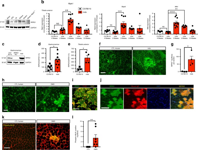
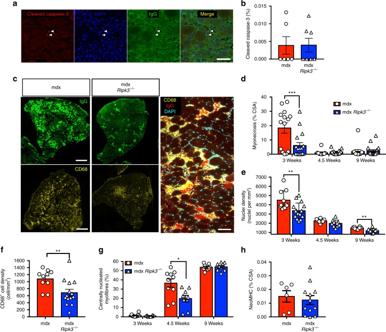
Duchenne muscular dystrophy (DMD) is a severe degenerative disorder caused by mutations in the dystrophin gene. Dystrophin-deficient muscles are characterised by progressive myofibre necrosis in which inflammation plays a deleterious role. However, the molecular mechanisms underlying inflammation-induced necrosis in muscle cells are unknown. Here we show that necroptosis is a mechanism underlying myofibre death in dystrophin-deficient muscle. RIPK1, RIPK3 and MLKL are upregulated in dystrophic mouse myofibres. In human DMD samples, there is strong immunoreactivity to RIPK3 and phospho-MLKL in myofibres. In vitro, TNFα can elicit necroptosis in C2C12 myoblasts, and RIPK3 overexpression sensitises myoblasts to undergo TNF-induced death. Furthermore, genetic ablation of Ripk3 in mdx mice reduces myofibre degeneration, inflammatory infiltrate, and muscle fibrosis, and eventually improves muscle function. These findings provide the first evidence of necroptotic cell death in a disease affecting skeletal muscle and identify RIPK3 as a key player in the degenerative process in dystrophin-deficient muscles.


Conflict of interest statement

The authors declare no competing interests.

Figures

Fig. 1
Fig. 1
Necroptosis is activated in mouse and human dystrophin-deficient muscles. a Immunoblot of RIPK3 protein expression in brain, extensor digitorum longus (EDL), tibialis anterior (TA), soleus, and gastrocnemius muscles of C57BL/6 and gastrocnemius from RIPK3 KO mouse. GAPDH was used as loading control. b Tibialis anterior muscles of 4-week-old C57BL/10, and 2-, 3-, 9-, and 13-week-old mdx mice were analysed for Ripk1, Ripk3, and Mlkl mRNA levels by quantitative PCR. Data were normalised to mouse Psma2 gene expression (n = 5 C57BL/10 and n = 5, 8, 10, 6, and 6 respectively, for 2-, 3-, 9-, and 13-week mdx TAs, Dunn’s multiple comparison test). c Protein extracts of C57BL/10, RIPK3 KO, and mdx gastrocnemius muscles were analysed by western blot for RIPK3 and GAPDH protein expression. Quantification of RIPK3 protein expression normalised to GAPDH in gastrocnemius (d) and TA (e) (n = 6 C57BL/10 and n = 9 mdx gastrocnemius; n = 3 C57BL/10 and n = 5 mdx TA, Student’s t-test). f Mouse gastrocnemius C57BL10 (left) and mdx (right) mice were immunolabelled with an antibody to RIPK3 (green). g Quantification of RIPK3-positive myofibres in gastrocnemius of C57BL10 (n = 3) and mdx mice (n = 4). h Representative transverse sections of quadriceps biopsies from a healthy non-dystrophic (CTL human) and a DMD patient, immunolabelled with an antibody to RIPK3 (green). i Representative image of immunostaining using antibodies to RIPK3 (green) and mouse IgG (red), a functional marker for membrane permeability in myofibres. j Representative image of immunostaining using antibodies to RIPK3 (green) and F4/80 (red), a marker for murine macrophages. k Confocal image of transverse cryosections of DMD quadriceps labelled with antibodies to human phospho-MLKL (green), human laminin α2 (red), and DAPI (blue). l Quantification of the number of p-MLKL-positive myofibres per field (n = 5 control human and n = 6 DMD). Data presented as the mean ± SEM. Scale bars, 100 µm. *P < 0.05, **P < 0.01, ***P < 0.001, ****P < 0.0001
Fig. 2
Fig. 2
RIPK3 upregulation facilitates TNFα-induced necroptosis in C2C12 myoblasts. a Immunoblot of RIPK3 expression in mouse embryonic fibroblast (MEF) cells and C2C12 myoblasts. b C2C12 myoblasts were exposed for 1, 2, or 3 h to either DMSO, mouse TNFα (100 ng/ml), TAK1 inhibitor (5Z)-7-Oxozeaenol (1 µM), and TNFα+TAK1 inhibitor (respectively T, O, and TO treatment) and cell survival was assessed by measuring ATP levels (CellTiter-Glo assay). Levels of ATP in DMSO-treated cells were defined as 100% survival. Data represented as the mean ± SEM of two independent experiments. c C2C12 myoblasts were challenged for 3 h with TNFα, (5Z)-7-Oxozeaenol, the pan-caspase inhibitor Z-VAD.fmk at 50 µM (Z), and the inhibitor of RIPK1 kinase activity Necrostatin-1s (Nec-1s) at 30 µM. Cell survival was assessed. Data presented as the mean ± SEM of three pooled independent experiments (n = 3, one-way ANOVA). d Same experimental conditions as in (c). Membrane permeability of treated cells was assessed using CytoTox-Glo assay after 3 h of treatment. Data represented as the mean ± SEM of three pooled independent experiments (n = 3, one-way ANOVA). e Cells were treated with TOZ for 3 h, with GSK’872, a RIPK3 inhibitor. Cell survival was measured. Data presented as the mean ± SEM of four pooled independent experiments (n = 4, one-way ANOVA). f Cells were treated as in e and membrane permeability was monitored by CytoTox-Glo assay. Data presented as the mean ± SEM of four pooled independent experiments (n = 4, one-way ANOVA). g Ripk3 or Mlkl mRNAs were knocked down using specific RNAi. Transfected C2C12 cells were treated with either TNFα alone or TOZ and cell survival was assessed. Representative of two independent experiments (five replicates per group). hi C2C12 myoblasts were challenged by TOZ with or without Butylated hydroxyanisole (BHA). Cell survival (h) and membrane permeability (i) were monitored. Data presented as the mean ± SEM of four pooled independent experiments (n = 4, one-way ANOVA). j C2C12 myoblasts were transfected with a GFP plasmid or a GFP-tagged Ripk3 plasmid. Transfected cells were stimulated and the survival was monitored (n = 4 pooled independent experiments, two-way ANOVA). Data shown as the mean ± SEM. *P < 0.05, **P < 0.01, ***P < 0.001, ****P < 0.0001
Fig. 3
Fig. 3
RIPK3 deficiency reduces the acute phase of myonecrosis in mdx mice. a Representative images of cleaved caspase-3 immunolabelling in 3-week-old mdxRipk3−/− TA. Scale bar, 50 µm. b Quantification of the percentage of cleaved caspase-3-positive nuclei. c Representative images of IgG uptake and CD68-positive cells in mdx (mdxRipk3+/+) and mdxRipk3−/− muscles. Scale bars, 500 µm (left panels) and 50 µm (right panel). d Quantification of the extent of myonecrosis with age in mdx and mdxRipk3−/− littermates. Animals were analysed at 3 weeks (n = 14 mdxRipk3+/+muscles, n = 17 mdxRipk3−/− muscles), 4.5 weeks (n = 10 mdx, n = 12 mdxRipk3−/−), and 9 weeks of age (n = 12 mdx, n = 18 mdxRipk3−/−). The extent of myonecrosis was defined as the area of IgG uptake in myofibres, and expressed as percentage of the cross-sectional area (%CSA). Two-way ANOVA, Sidak’s multiple comparisons test. e Quantification of nuclear density in TA muscles at 3 weeks (n = 10 mdx, n = 16 mdxRipk3−/−), 4.5 weeks, and 9 weeks (n = 8 mdx, n = 10 mdxRipk3−/−). f Quantification of CD68-positive cell infiltration in TA of 3-week-old mice (n = 10 mdxRipk3+/+,n = 12 mdxRipk3−/− muscles, Student’s t-test). f Quantification of neoMHC staining in 4.5-week-old mice. Data expressed as percentage of cross-section area (n = 6 mdxRipk3+/+ and n = 12 mdxRipk3−/− TA). g Quantification of the percentage of centrally nucleated fibres (n = 8 mdxRipk3+/+ and n = 9 mdxRipk3−/− TA). h Quantification of NeoMHC-positive myofibres at 4.5 weeks (% of CSA). Data presented as the mean ± SEM. *P < 0.05, **P < 0.01, ***P < 0.001
Fig. 4
Fig. 4
RIPK3 deficiency ameliorates muscle phenotype in adult mice. a Representative image of extracellular matrix deposition in TA muscles of 9-week-old mice. Extracellular matrix was stained with antibodies to laminin and collagen VI together (red). Scale bar, 500 µm. b Quantification of extracellular matrix deposition (fibrosis) in TA of 9-week-old mice. Total extracellular matrix area was expressed as percentage of cross-sectional area (n = 7 mdx, n = 11 mdxRipk3−/−, Student’s t-test). c Quantification of serum CK in 3-month-old mdx mice (n = 13 mdx, n = 17 mdxRipk3−/−, Mann–Whitney test). d Quantification of grip strength and e hanging performance (expressed as the ratio: maximum hanging time (in s)/body weight; n = 8 mdx, n = 12 mdxRipk3−/−, Mann–Whitney test), f Quantification of spontaneous activity of mice in 12 h time. Data presented as the mean ± SEM. *P < 0.05, **P < 0.01

References

    1. Gilbert RK, Hawk WA. The incidence of necrosis of muscle fibers in Duchenne type muscular dystrophy. Am. J. Pathol. 1963;43:107–122. - PMC - PubMed
    1. Moat SJ, Bradley DM, Salmon R, Clarke A, Hartley L. Newborn bloodspot screening for Duchenne muscular dystrophy: 21 years experience in Wales (UK) Eur. J. Hum. Genet. 2013;21:1049–1053. doi: 10.1038/ejhg.2012.301. - DOI - PMC - PubMed
    1. Matsuda R, Nishikawa A, Tanaka H. Visualization of dystrophic muscle fibers in mdx mouse by vital staining with Evans blue: evidence of apoptosis in dystrophin-deficient muscle. J. Biochem. 1995;118:959–964. doi: 10.1093/jb/118.5.959. - DOI - PubMed
    1. Tidball JG, Albrecht DE, Lokensgard BE, Spencer MJ. Apoptosis precedes necrosis of dystrophin-deficient muscle. J. Cell Sci. 1995;108(Pt 6):2197–2204. - PubMed
    1. Sandri M, Carraro U. Apoptosis of skeletal muscles during development and disease. Int. J. Biochem. Cell Biol. 1999;31:1373–1390. doi: 10.1016/S1357-2725(99)00063-1. - DOI - PubMed

Publication types

Substances