. 2018 Sep 7;8(1):13437.
doi: 10.1038/s41598-018-31542-y.

Neuronal microRNA regulation in Experimental Autoimmune Encephalomyelitis

Affiliations

Neuronal microRNA regulation in Experimental Autoimmune Encephalomyelitis

Camille A Juźwik et al. Sci Rep. .

Abstract

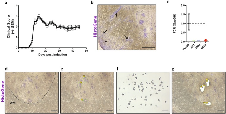
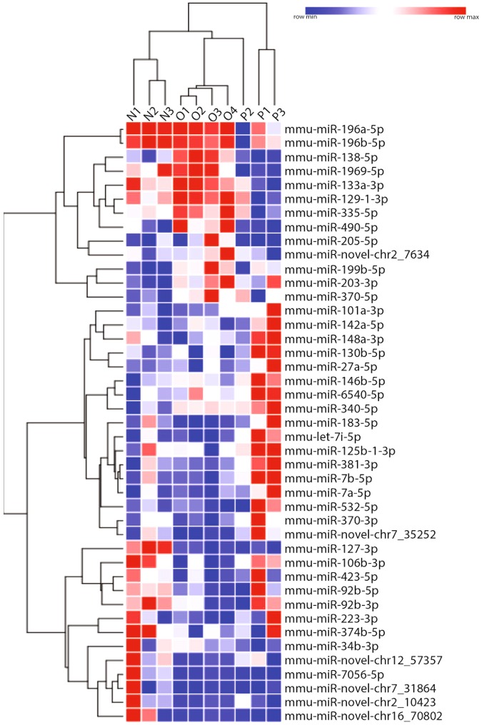
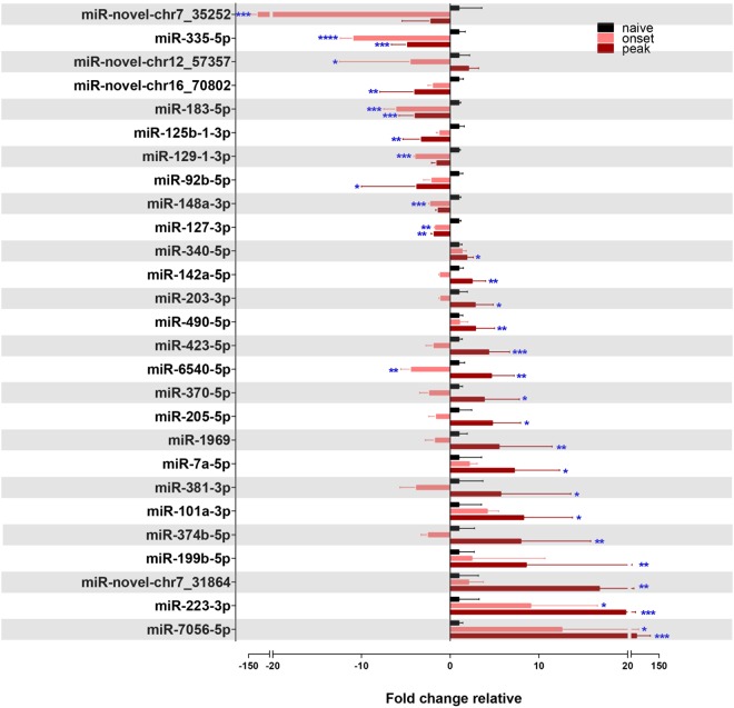
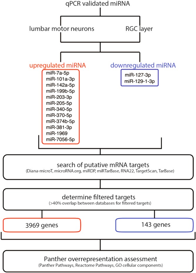
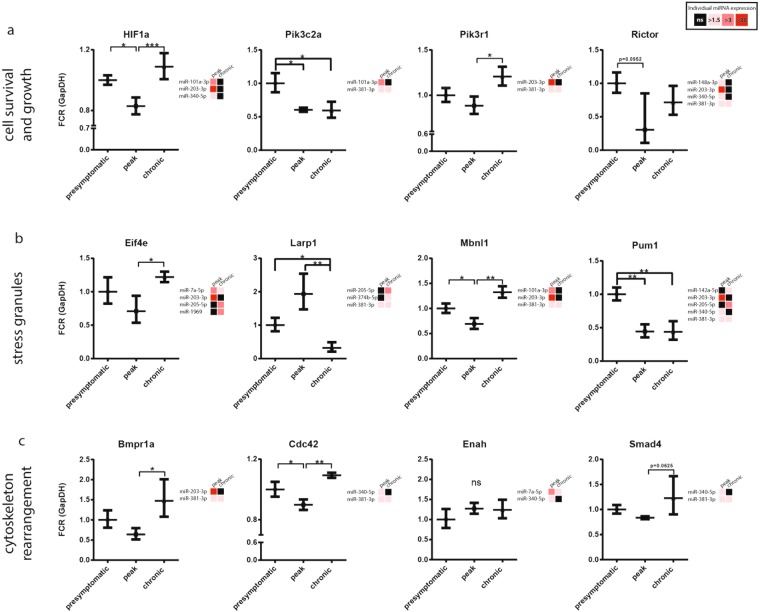
Multiple sclerosis (MS) is an autoimmune, neurodegenerative disease but the molecular mechanisms underlying neurodegenerative aspects of the disease are poorly understood. microRNAs (miRNAs) are powerful regulators of gene expression that regulate numerous mRNAs simultaneously and can thus regulate programs of gene expression. Here, we describe miRNA expression in neurons captured from mice subjected to experimental autoimmune encephalomyelitis (EAE), a model of central nervous system (CNS) inflammation. Lumbar motor neurons and retinal neurons were laser captured from EAE mice and miRNA expression was assessed by next-generation sequencing and validated by qPCR. We describe 14 miRNAs that are differentially regulated in both neuronal subtypes and determine putative mRNA targets though in silico analysis. Several upregulated neuronal miRNAs are predicted to target pathways that could mediate repair and regeneration during EAE. This work identifies miRNAs that are affected by inflammation and suggests novel candidates that may be targeted to improve neuroprotection in the context of pathological inflammation.


Conflict of interest statement

Dr. Bar-Or has participated as a speaker in meetings sponsored by and received consulting fees and/or grant support from: Atara Biotherapeutics, Biogen Idec, Celgene/Receptos, Genentech/Roche, GlaxoSmithKline, MAPI, Medimmune, Merck/EMD Serono, Novartis, Sanofi-Genzyme.

Figures

Figure 1
Figure 1
Laser captured lumbar motor neurons from EAE and control naive mice. (a) Clinical scores of mice immunized with MOG35–50 peptide. Animals of interest were taken at onset (score 0.5 to 1) and peak (score 3–3.5) of disease. Curve represents a cohort of 38 immunized animals, with representative animals taken at onset and peak. (b) Representative EAE spinal cord section from an animal at peak disease (score 3–3.5) stained with HistoGene and displaying immune cell infiltrates (arrows) and areas of demyelination (*) (scale bar at 100 um). (c) qPCR of LCM motor neuron tissue for microglia/macrophages (Aif1), immune cells (Cd3e), and astrocytes (Gfap) relative to neuronal RNA expression (Tubb3). FCR, Fold Change Range. (d–g) Micrographs of the laser capture microdissection flow-through. Sections of frozen mouse spinal cord on PEN membrane slides stained with HistoGene. Area above the dotted line is the dorsal horn and the area below is the white matter, WM. (d) The tissue surrounding the lumbar motor neurons was traced and ablated loosening the neuron of interest. (e) The neuron was then catapulted into an adhesive cap (f) leaving a void on the slide. (g) Scale bar; 50 um (d,e,g) 250 um (f).
Figure 2
Figure 2
Heat Map summary of significantly regulated miRNA as identified by miR-Seq in EAE. miRNA and animals (N = naive, O = onset, P = peak) are hierarchically clustered by Euclidean distance using rlog transformed counts. Blue data represents low expression of that microRNA within its own row, and red indicates high expression. 997 miRNAs were identified by miR-Seq, 43 of these were identified as significantly regulated (p < 0.05); 6 of which are novel miRNAs.
Figure 3
Figure 3
Differentially regulated miRNAs in the lumbar motor neurons of EAE mice over the course of the disease. Taqman MicroRNA Assay (qPCR) validation of miR-Seq identified differentially regulated miRNAs in the lumbar motor neurons of EAE mice over the course of the disease, normalized to endogenous control snoRNA202 for each individual miRNA and depicted as fold change relative to normalized naive levels. ****p < 0.0001, ***p < 0.001, p** < 0.01, *p < 0.05 (n = 3–6, one-way ANOVA, p < 0.05, Dunnett’s multiple comparisons test).
Figure 4
Figure 4
Profiling miRNA expression in the RGC layer of EAE mice. (a) Representative retinal sections stained with HistoGene following LCM (ROI = region of interest, RGC = retinal ganglion cell layer, IPL = inner plexiform layer, INL = inner nuclear layer, OPL = outer plexiform layer, ONL = outer nuclear layer), 50 um scale bar. (b) qPCR of LCM RGC layer tissue for microglia/macrophages (Aif1), immune cells (Cd3e), and astrocytes (Gfap) relative to neuronal RNA expression (Tubb3)., FCR, Fold Change Range. (c) Taqman MicroRNA Assay (qPCR) of miRNAs in the RGC layer of EAE mice over the course of the disease, normalized to endogenous control snoRNA202 for each individual miRNA and depicted as fold change relative to normalized presymptomatic levels. ****p < 0.0001, ***p < 0.001, p** < 0.01, *p < 0.05 (n = 3–8, one-way ANOVA, p < 0.05, Dunnett’s multiple comparisons test).
Figure 5
Figure 5
Flow-through of the in silico assessment for putative mRNA targets of the differentially regulated miRNAs in the lumbar motor neurons and RGC layer of EAE mice.
Figure 6
Figure 6
The upregulated miRNAs of neurons during EAE potentially block neuroprotective responses to inflammation. Upregulated miRNAs block axon guidance cues, PIP synthesis, CD28 co-stimulation, HIF activation, KIT signaling, and transcriptional and post-transcriptional regulation pathways, as determined by an in silico approach. PI3K/Akt signaling for potential cell survival is blocked by the inhibition of PIP synthesis, HIF activation and KIT signaling. Inhibition of KIT signaling, along with CD28 co-stimulation, prevents any potential dampening of CNS inflammation. Inhibition of several axon guidance cues occurs via the inhibition of signaling through Netrin-1, Slit/Robo and BMP. Finally, inhibition of PKMT histone lysine methylation, CCR4-NOT complex assembly, and stress granule formation converge on many levels of transcriptional and post-transcriptional regulation.
Figure 7
Figure 7
Expression of predicted miRNA gene targets as identified by in silico analysis in the RGC layer. Right-hand side of every qPCR profile for target genes is their respective targeting miRNA expression profiles with fold upregulation relative to presymptomatic levels. Black, not significant (ns). Genes are organized by their physiological roles, specifically (a) cell survival and growth (b) stress granule formation, and (c) cytoskeleton rearrangement. Gene expression was normalized to endogenous GapDH, FCR = Fold Change Range. ***p < 0.001, **p < 0.01, *p < 0.05 (n = 3–6, one-way ANOVA, p < 0.05, Tukey’s multiple comparisons test).

References

    1. Frohman EM, Racke MK, Raine CS. Multiple sclerosis–the plaque and its pathogenesis. The New England journal of medicine. 2006;354:942–955. doi: 10.1056/NEJMra052130. - DOI - PubMed
    1. Dutta, R. & Trapp, B. D. Pathogenesis of axonal and neuronal damage in multiple sclerosis. Neurology 68, S22–31; discussion S43–54 (2007). - PubMed
    1. Fisher JB, et al. Relation of visual function to retinal nerve fiber layer thickness in multiple sclerosis. Ophthalmology. 2006;113:324–332. doi: 10.1016/j.ophtha.2005.10.040. - DOI - PubMed
    1. Ferguson B, Matyszak MK, Esiri MM, Perry VH. Axonal damage in acute multiple sclerosis lesions. Brain. 1997;120(Pt 3):393–399. doi: 10.1093/brain/120.3.393. - DOI - PubMed
    1. Peterson JW, Bo L, Mork S, Chang A, Trapp BD. Transected neurites, apoptotic neurons, and reduced inflammation in cortical multiple sclerosis lesions. Ann Neurol. 2001;50:389–400. doi: 10.1002/ana.1123. - DOI - PubMed

Publication types

MeSH terms

Grants and funding

LinkOut - more resources