. 2018 Nov;109(11):3643-3656.
doi: 10.1111/cas.13795. Epub 2018 Oct 5.

Girdin/GIV regulates collective cancer cell migration by controlling cell adhesion and cytoskeletal organization

Affiliations

Girdin/GIV regulates collective cancer cell migration by controlling cell adhesion and cytoskeletal organization

Xiaoze Wang et al. Cancer Sci. 2018 Nov.

Abstract

Pathological observations show that cancer cells frequently invade the surrounding stroma in collective groups rather than through single cell migration. Here, we studied the role of the actin-binding protein Girdin, a specific regulator of collective migration of neuroblasts in the brain, in collective cancer cell migration. We found that Girdin was essential for the collective migration of the skin cancer cell line A431 on collagen gels as well as their fibroblast-led collective invasion in an organotypic culture model. We provide evidence that Girdin binds to β-catenin that plays important roles in the Wnt signaling pathway and in E-cadherin-mediated cell-cell adhesion. Girdin-depleted cells displayed scattering and impaired E-cadherin-specific cell-cell adhesion. Importantly, Girdin depletion led to impaired cytoskeletal association of the β-catenin complex, which was accompanied by changes in the supracellular actin cytoskeletal organization of cancer cell cohorts on collagen gels. Although the underlying mechanism is unclear, this observation is consistent with the established role of the actin cytoskeletal system and cell-cell adhesion in the collective behavior of cells. Finally, we showed the correlation of the expression of Girdin with that of the components of the E-cadherin complex and the differentiation of human skin cancer. Collectively, our results suggest that Girdin is an important modulator of the collective behavior of cancer cells.

Keywords: Girdin; actin cytoskeleton; cell adhesion; collective invasion; collective migration.


Figures

Figure 1
Figure 1
Girdin regulates the collective migration of A431 cells on collagen gels. A, The behavior of single cells on collagen gels is different from that on conventional plastic dishes. The directionality of migrating single cells was calculated as the ratio (d/D), that is, the distance between the starting and ending points (d) divided by the actual trajectory (D). Ten and five dishes were evaluated for the collagen gel group and plastic dish group, respectively, and 25 cells in each dish were manually tracked, followed by quantification. B, Representative images of A431 cell groups cultured on plastic dishes (upper panel) and a collagen gel (lower panel). Note that the cell groups (dotted circles) undergo proliferation without movement on plastic dishes, whereas those seeded on the collagen gel collectively migrate with directionality. Arrows indicate the direction of the movement of the cell group. See also Movies S1 and S2. C, Schematic illustration showing the measurement of collective migration of A431 cells cultured on a collagen gel. We focused on one single cell on the edge of migrating cell groups and tracked its trajectory by tracing the nuclear centroid. D, shRNA‐mediated depletion of Girdin in A431 cells. MW, molecular weight. E‐G, Representative images of control and Girdin‐depleted A431 cell groups cultured on a collagen gel. Time interval between each panel is 2 hours. Note that the control cell group underwent collective directional migration, whereas the Girdin‐depleted cells tended to remain in place. Shown in (F) are the representative paths of the migration of control shRNA‐ (red) and Girdin shRNA (1; green)‐transduced cell groups, as determined by tracing the nuclear centroid over a period of 5 hours (n = 25 for each group). The directionality of migrating cell groups was quantified and shown in G (n = 50 for each group)
Figure 2
Figure 2
Interaction of Girdin with the β‐catenin complex. A, B, Immunoprecipitation (IP) with anti‐Girdin antibody showed that Girdin interacts with β‐ and α‐catenins in A431 cells (A). Interaction between Girdin and catenins was also shown by reciprocal IP with anti‐β‐catenin antibody (B). TCL, total cell lysates. C, D, Immunofluorescence staining showed the colocalization of Girdin (green) with α‐ and β‐catenins (red) in A431 cells seeded on plastic dishes (C) and collagen gels (D). E, Domain structures and interacting proteins of human Girdin and β‐catenin. Fragments and domains of Girdin and β‐catenin used in the study are shown. The domains responsible for the interaction are shown in red. F, Mapping of interacting domains of Girdin and β‐catenin. β‐Catenin‐binding site maps to the C‐terminal (CT) domain of Girdin. Lysates from 293FT cells transfected with the Girdin fragments fused with GFP were immunoprecipitated with anti‐GFP antibody, followed by western blot analyses using β‐ and α‐catenin antibodies. Bound catenins are indicated by asterisks. G, Girdin‐binding site maps to the N‐terminal (NT) domain of β‐catenin. Lysates from 293FT cells transfected with the Flag tag‐fused Girdin CT domain and the β‐catenin fragments fused with GST were precipitated with glutathione‐Sepharose beads, followed by western blot analyses using anti‐Flag antibody. Bound Flag‐Girdin CT is indicated by asterisks. H, I, Direct interaction of Girdin and β‐catenin. Coomassie brilliant blue staining showing recombinant GST, GST‐fused β‐ and α‐catenins that were expressed and purified from the Escherichia coli expression system (H). Purified recombinant Girdin CT domain was incubated with the recombinant catenins fused with GST (60‐90 pmol) for 1 hour at 4°C, followed by precipitation with glutathione‐Sepharose beads and western blot analysis (I). Girdin CT domain that bound to GST‐catenins is indicated by asterisks. MW, molecular weight
Figure 3
Figure 3
Girdin controls the strength of cell‐cell adhesion. A, Girdin localized at cell‐cell contacts. Its colocalization with β‐catenin was not evident in immature cell‐cell adhesion of A431 cells sparsely plated on dishes (left), whereas it was clearly observed in confluent cells with mature cell‐cell adhesion (right). B, C, HeLa cells depleted of Girdin showed morphology of scattered cells. D, Efficiency of siRNA‐mediated knockdown of Girdin, E‐cadherin, and α‐ and β‐catenins in A431 cells was shown by western blot analyses using the indicated antibodies. E, F, A431 cells transfected with the indicated siRNA were treated with trypsin for 30 minutes in the presence of either 0.1 mmol/L Ca2+ (TC) or 1 mmol/L EDTA (TE) at 37°C. The cells were dissociated by pipetting, and the number of particles was counted. Cells of each group were seeded in three 6‐cm dishes, followed by counting the numbers of all particles in the dishes and quantification. Representative images are shown in (E). The extent of cell dissociation was represented by the index TC/TE, where TC and TE are the total particle numbers after the TC and TE treatment, respectively (F). The TC/TE ratio represents the inverse strength of cadherin activity. G, H, shRNA‐mediated Girdin depletion decreased cadherin activity in HeLa cells, which is shown by high TC/TE values in the cell dissociation assay
Figure 4
Figure 4
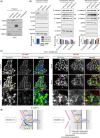
Girdin participates in the cytoskeletal association of the E‐cadherin/catenin complex and cytoskeletal organization. A, Representative data showing successful fractionation of various subcellular fractions of A431 cells. B, Cytosolic, cytoskeletal, and membrane fractions from A431 cells transfected with control or Girdin siRNA were examined by western blot analyses using the indicated antibodies. Lower panel, expression levels of the proteins in each fraction were quantified and presented as arbitrary unit (AU). EGFR, epidermal growth factor receptor; HSP70, heat shock protein 70. C, Control cells (left panel) and Girdin‐depleted cells (right panel) were stained by β‐catenin (red) and phalloidin to visualize actin filaments (green). Yellow arrowheads denote filopodia formation found in the edges and cell‐cell contact sites at the basal planes of control cell groups, whereas filopodia formation was weak or disrupted in Girdin‐depleted cells (magenta arrowheads). D, Schematic illustration of the speculated function of Girdin at cell‐cell adhesions. Girdin mediates the link of β‐catenin and the actin cytoskeleton (left panel), which is lost by the absence of Girdin accompanied by dysregulated cytoskeletal organization (right panel). A previous study showed that Girdin interaction with Dynamin is essential for E‐cadherin endocytosis, which might also be involved in collective cell migration. CT, C‐terminal domain; NT, N‐terminal domain
Figure 5
Figure 5
Effect of overexpression of the β‐catenin N‐terminal (NT) domain on collective migration of A431 cells. A, Inhibition of endogenous Girdin/β‐catenin interaction by expression of the β‐catenin NT domain. Lysates from A431 cells transfected with the indicated combination of expression plasmids were immunoprecipitated (IP) with anti‐Girdin antibody. After washing, bound proteins were detected by western blot analyses using the indicated antibodies. β‐Catenin precipitated by Girdin antibody is shown by asterisk. B, A431 cells were transfected with GFP (as a fill) and either GST or GST‐β‐catenin NT at the ratio of 1:4, and were sorted for GFP‐positive cells. They were cultured on collagen gels and assessed by tracking of single cells on the edge of migrating cell groups (≥5 cells). C, Representative images of GST and GST‐β‐catenin NT‐transduced A431 cell groups cultured on collagen gels. Time interval between each panel is 2 hours. D, Quantification of the directionality of migrating cell groups (n = 18 for each group)
Figure 6
Figure 6
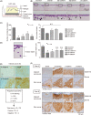
Girdin is essential for collective invasion of cancer cells. A, Illustration showing the experimental setup to recapitulate the collective invasion of cancer cells in vitro. CAF, cancer‐associated fibroblast. B, C, Depletion of Girdin and the components of the E‐cadherin complex impedes the collective invasion of A431 cells. Representative H&E‐stained images of sections through Matrigel invaded by A431 cells. Boxed regions are magnified in lower panels, where arrows denote the groups of cells collectively invading into Matrigel. In (C), the numbers of cell groups (clusters; ≥3 cells) invading into Matrigel and cell numbers in each cluster were counted and quantified (n = 3). D, Depth of invasion for each cluster found in each group was calculated and quantified (n = 3). E, Images of representative Girdin staining intensity for each intensity score (IS; 0‐3). Cases with total scores (TS), which represents the sum of IS and proportion scores (PS), of >3 were considered positive. F, Representative images of immunohistochemical staining for Girdin and the components of the E‐cadherin/catenin complex in cases A and B of invasive squamous cell carcinoma of the skin. Invasive lesions of the tumors (lower panels) and the adjacent normal skin (upper panels) are shown. Note that Girdin expression in the carcinomas is more evident than in normal skin

Similar articles

Cited by

References

    1. Chaffer CL, Weinberg RA. A perspective on cancer cell metastasis. Science. 2011;331:1559‐1564. - PubMed
    1. Yang J, Weinberg RA. Epithelial‐mesenchymal transition: at the crossroads of development and tumor metastasis. Dev Cell. 2008;14:818‐829. - PubMed
    1. Yilmaz M, Christofori G. EMT, the cytoskeleton, and cancer cell invasion. Cancer Metastasis Rev. 2009;28:15‐33. - PubMed
    1. Lamouille S, Xu J, Derynck R. Molecular mechanisms of epithelial‐mesenchymal transition. Nat Rev Mol Cell Biol. 2014;15:1781‐1796. - PMC - PubMed
    1. Ridley AJ. Rho GTPase signalling in cell migration. Curr Opin Cell Biol. 2015;36:103‐112. - PMC - PubMed