

In Vivo Confocal Microscopy Shows Alterations in Nerve Density and Dendritiform Cell Density in Fuchs' Endothelial Corneal Dystrophy
- PMID: 30194928
- PMCID: PMC6420962
- DOI: 10.1016/j.ajo.2018.08.040
In Vivo Confocal Microscopy Shows Alterations in Nerve Density and Dendritiform Cell Density in Fuchs' Endothelial Corneal Dystrophy
Abstract
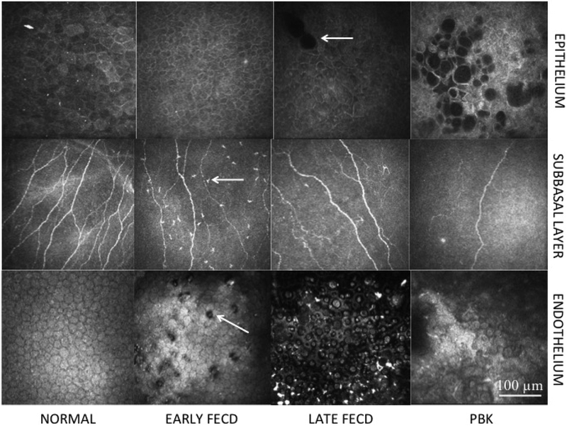
Purpose: To evaluate corneal nerve and immune cell alterations in Fuchs' endothelial corneal dystrophy (FECD) and pseudophakic bullous keratopathy (PBK) by laser in vivo confocal microscopy (IVCM) as correlated to corneal sensation and endothelial cell loss.
Design: Prospective, cross-sectional, controlled study.
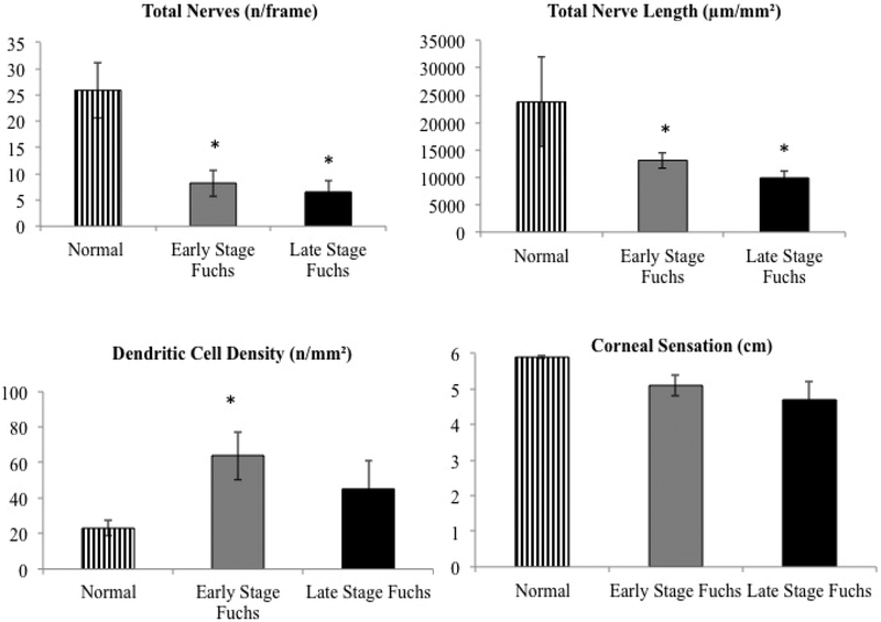
Methods: Thirty-three eyes with FECD were compared to 13 eyes with PBK and 17 normal age-matched control eyes at a tertiary referral center. FECD was classified into early (without edema) and late stage (with edema). Corneal IVCM and esthesiometry were performed. Corneal nerve and immune dendritiform cell (DC) alterations were evaluated and correlated to clinical parameters.
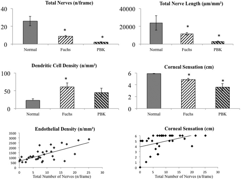
Results: FECD and PBK eyes showed significantly (P = .001) diminished total nerve length (11.5 ± 1.3 and 2.9 ± 0.7 mm/mm2) and number (8.8 ± 1.1 and 2.2 ± 0.4 n/frame), compared to controls (23.3 ± 8.1 mm/mm2 and 25.9 ± 1.3 n/frame). Decreased nerves corresponded to diminished sensation in FECD (4.9 ± 0.2 cm; R = 0.32; P = .045), compared to controls (5.9 ± 0.04 cm). Early- and late-stage FECD showed significantly reduced total nerve length (13.1 ± 1.4 and 9.9 ± 1.2 mm/mm2, respectively) and number (8.2 ± 2.5 and 6.5 ± 2.1 n/frame), compared to controls (P < .001). DC density was significantly increased in FECD (57.8 ± 10.4 cells/mm2; P = .01), but not in PBK (47.7 ± 11.6 cells/mm2; P = .60) compared to controls (22.5 ± 4.5 cells/mm2). A subset of early FECD patients (7/22) demonstrated very high DC density (>100/mm2).
Conclusion: IVCM demonstrates profound diminishment of subbasal corneal nerves in early- and late-stage FECD and in PBK, correlating to decreased sensation. Increased DC density in early FECD demonstrates potential subclinical inflammation. The data suggest that reduction in subbasal nerves and increased immune activation may play a role in the pathophysiology of FECD.
Copyright © 2018 Elsevier Inc. All rights reserved.
Conflict of interest statement
b. Financial Disclosures: No financial disclosures for any authors
Figures
Comment in
-
Bindehaut- und Hornhauterkrankungen.Ophthalmologe. 2019 Nov;116(11):1006-1007. doi: 10.1007/s00347-019-00958-w. Ophthalmologe. 2019. PMID: 31696286 German. No abstract available.
References
-
- EBAA 2012. Statistical Report. http://www.restoresight.org/wp-content/uploads/2014/04/2013_Statistical_....
-
- Ghosheh FR, Cremona FA, Rapuano CJ, et al. Trends in penetrating keratoplasty in the United States 1980–2005. Int Ophthalmol. 2008;28(3):147–153. - PubMed
-
- Fuchs E Dystrophia epithelias corneae. Graefes Arch Klin Exp Ophthalmol. 1910;76:478–508.
-
- Moller HU, Weiss JS. IC3D classification of corneal dystrophies. Dev Ophthalmol. 2011;48:1–8. - PubMed
Publication types
MeSH terms
Grants and funding
LinkOut - more resources
Full Text Sources
Other Literature Sources
