. 2018 Nov:74:176-185.
doi: 10.1016/j.bbi.2018.09.004. Epub 2018 Sep 5.

The β2-adrenergic receptor controls inflammation by driving rapid IL-10 secretion

Affiliations

The β2-adrenergic receptor controls inflammation by driving rapid IL-10 secretion

Didem Ağaç et al. Brain Behav Immun. 2018 Nov.

Abstract

The mammalian nervous system communicates important information about the environment to the immune system, but the underlying mechanisms are largely unknown. Secondary lymphoid organs are highly innervated by sympathetic neurons that secrete norepinephrine (NE) as the primary neurotransmitter. Immune cells express adrenergic receptors, enabling the sympathetic nervous system to directly control immune function. NE is a potent immunosuppressive factor and markedly inhibits TNF-α secretion from innate cells in response to lipopolysaccharide (LPS). In this study, we demonstrate that NE blocks the secretion of a variety of proinflammatory cytokines by rapidly inducing IL-10 secretion from innate cells in response to multiple Toll-like receptor (TLR) signals. NE mediated these effects exclusively through the β2-adrenergic receptor (ADRB2). Consequently, Adrb2-/- animals were more susceptible to L. monocytogenes infection and to intestinal inflammation in a dextran sodium sulfate (DSS) model of colitis. Further, Adrb2-/- animals rapidly succumbed to endotoxemia in response to a sub-lethal LPS challenge and exhibited elevated serum levels of TNF-α and reduced IL-10. LPS-mediated lethality in WT animals was rescued by administering a β 2-specific agonist and in Adrb2-/- animals by exogenous IL-10. These findings reveal a critical role for ADRB2 signaling in controlling inflammation through the rapid induction of IL-10. Our findings provide a fundamental insight into how the sympathetic nervous system controls a critical facet of immune function through ADRB2 signaling.

Keywords: Adrenergic receptor; Inflammation; Interleukin-10; Macrophage; Norepinephrine; Sepsis.


Conflict of interest statement

COMPETING INTERESTS

The authors declare no competing interests.

Figures

Figure 1.
Figure 1.. NE suppresses TLR-induced pro-inflammatory cytokine secretion in bone marrow-derived macrophages.
(A) BM-DMs from Balb/cJ mice were stimulated with one of the following TLR agonists: 100 ng/mL Pam3CSK4, 10 μg/mL poly l:C, 100 ng/mL LPS, or 1 ng/mL R837 in the absence or presence of 5 μΜ NE for 2, 8 and 48 hours. Error bars represent the standard error of the mean for n=2 mice. (B) BM-DMs from C57BI/6J mouse were stimulated in the presence or absence of 5 μΜ NE and 1 μΜ CpG for 2–48 hours. Cytokines in the supernatant were quantified using Mouse Pro- inflammatory 7-Plex Tissue Culture Kit from Mesoscale Discovery.
Figure 2.
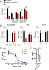
Figure 2.. NE suppresses TLR-induced pro-inflammatory cytokine secretion through the ADRB2.
(A) BM-DMs from C57BI/6J mouse were stimulated with one of the following adrenergic receptor antagonists: 1 μΜ phentolamine (aAR antagonist), 1 μΜ nadolol (PAR antagonist), 100 nM atenolol (P1AR antagonist), or 100 nM ICI118,551 (P2AR antagonist) in the presence or absence of 5 μΜ NE and 1 μΜ CpG for 24 hours. TNF-α in the supernatants was quantified by ELISA. (B) WT and Adrb2−/− BM-DMs were stimulated with different TLR ligands in the presence or absence of norepinephrine for 24 hours. Supernatants were harvested and TNF-α in the supernatant was quantified by ELISA. Error bars represent the standard error of the mean for n=3 mice per strain. (C) BM-DMs were stimulated with 1 μΜ CpG and increasing concentrations of albuterol for 2, 8 and 48 hours. Error bars represent the standard error of the mean for n=3 mice per strain. (D) Whole blood from healthy adult donors were stimulated with 10 μg/mL LPS in the presence or absence of 50 nM albuterol for 4 hours. Plasma TNF-α was assessed by ELISA. The data represents 6 donors. Two-tailed paired t-test was conducted for (D), p<0.05.
Figure 3.
Figure 3.. Engagement of adrenergic receptors induce rapid IL-10 production.
(A) BM-DMs from C57BI/6J mouse were stimulated in the presence or absence of 5 μΜ NE and 1 μΜ CpG for 2–48 hours. (B) Adrb2- sufficient and -deficient BM-DMs were stimulated with 100 ng/mL LPS in the presence or absence of norepinephrine for 24 hours. Supernatants were harvested and IL-10 in the supernatant was quantified using Mouse Pro-inflammatory 7-Plex Tissue Culture Kit from Mesoscale Discovery. Error bars represent the standard error of the mean for n=3 mice per strain. (C) BM-DMs were stimulated with one of the following TLR agonists: 100 ng/mL Pam3CSK4, 10 pg/mL poly l:C, 100 ng/mL LPS, 1 pg/mL R837, or 1 μΜ CpG for 2 hours in the absence (black bars) or presence (red bars) of 5 μΜ NE. The cytokines released to the supernatant were quantified using Mouse Pro-inflammatory 7-Plex Tissue Culture Kit from Mesoscale Discovery. (D) Human monocyte-derived macrophages were stimulated with 10 pg/mL LPS in the presence or absence of 5 μΜ NE for 2 hours. Secreted TNFa and IL-10 were quantified by ELISA. The data represents 4 donors. **p<0.001 by Bonferroni post-hoc test following two-way ANOVA (B) and p<0.05 by two- tailed paired t-test (D).
Figure 4.
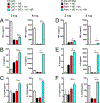
Figure 4.. NE-mediate regulation of cytokine expression in II–10−/− macrophages.
.WT and Il10−/− BM-DMs were stimulated with 1 μΜ CpG (A-C) or 100 ng/mL LPS (D-F) in the presence or absence of 5 μΜ NE for 2, 4, 8, and 24 hours. Cytokines in the supernatant were quantified using Mouse Pro-inflammatory 7-Plex Tissue Culture Kit from Mesoscale Discovery. Bar graphs on the left of each panel are data from the 2 hr timpoint that are included in the kinetic data displayed in the line graphs on the left of each panel. Error bars represent the standard error of the mean for n=2–3 mice per strain. p<0.05 by two-tailed paired t-test within each strain.
Figure 5.
Figure 5.. The role of the IL-10R in NE-mediated suppression of the pro-inflammatory cytokine secretion.
BM-DMs from Balb/cJ mouse were pre-treated with 10 pg/mL anti-CD210 (IL-10R) antibody or isotype control for 30 minutes. BM-DMs were stimulated with 1 μΜ CpG (A-C) or 100 ng/mL Pam3CSK4 (D-F) in the absence or presence of 5 μΜ NE for 2 and 8 hours. Cytokines were quantified with Mouse Pro- inflammatory 7-Plex Tissue Culture Kit from Mesoscale Discovery. p<0.05 by Tukey’s post-hoc test following ordinary one-way ANOVA across treatment groups.
Figure 6.
Figure 6.. Adrb2 signaling controls innate responses to bacterial infection.
(A) WT and Adrb2−/− mice were challenged with a lethal dose of LM (LM-OVA, 277K CFU/mouse) intravenously (i.v.) and the survival of the animals were monitored for 7 days. (n=10–11 mice per strain). (B) WT and Adrb2−/− mice were challenged with sub- lethal dose of LM-OVA i.v. and spleen weights were assessed during infection as a proxy for inflammation (n=7–16 mice per strain). (C) WT and Adrb2−/− mice were challenged with sub-lethal LM-OVA and were administered 40 pg Salmeterol at days 0, 1 and 2 of infection intraperitoneally. Spleen weights were assessed day 7 postinfection (n=12–16 mice per condition). p=0.0422 by log-rank test (A), p<0.05 by Bonferroni post-hoc test following two-way ANOVA (B), p<0.05 by Tukey’s post-hoc test following two-way ANOVA (C).
Figure 7.
Figure 7.. Adrb2-deficient animals are susceptible to DSS colitis.
WT and Adrb2−/− mice were given either water or 3% DSS diluted in their drinking water for 7 days, and body weights were measured for each animal during the course of the experiment. (A) The percent of starting body was calculated through d7, at which time animals were sacrificed to measure colon length (B) and histopathology. (C) Linear regression analysis of colon length versus histopathology scoring of colon inflammation. (D) Representative histopathology images of colons from two separate mice from either the WT or Adrb2−/− groups. n=7–8 mice per strain. p<0.05 by Bonferroni post-hoc test following two-way ANOVA.
Figure 8.
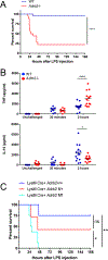
Figure 8.. Adrb2-deficient animals are susceptible to endotoxemia.
WT and Adrb2−/− mice were challenged with 300 pg LPS i.p. (A) Survival of the animals was monitored for 7 days (n=18–20 mice), and (B) serum cytokines were assessed in separate cohorts of animals by ELISA (n=9–14 mice). (C) LysM-Cre+ WT, Adrb2fx/+ and Adrb2fx/fx mice were challenged with 250 pg LPS i.p. and the survival of the animals were monitored for 7 days (n=4–8 mice). p<0.05 by log-rank test (A and C), p<0.05 by Bonferroni post-hoc test following two-way ANOVA.
Figure 9.
Figure 9.. ADRB2 signaling controls IL-10 production in vivo.
(A) WT and Adrb2−/− mice were challenged i.p. with 200–250 μg LPS. A cohort of Adrb2−/− were administered 1 μg recombinant murine IL-10 i.p. immediately after LPS challenge. The survival of the animals was monitored for 7 days (n=13–14 mice per condition) (B) WT Balb/c mice were challenged with 750 pg LPS, in the presence of 40 pg salmeterol (or vehicle control) and/or 200 pg anti CD210 (IL-10R) antibody (or isotype control). The survival of animals was monitored for 7 days. (n=17–20 mice per treatment group) (C) WT Balb/c mice were challenged with 300 pg LPS in the absence or presence of 40 pg salmeterol. Blood was harvested at indicated time points and serum cytokines were quantified by ELISA. (n=3–10 mice per time point). p<0.05 by log-rank test (A and B), p<0.05 by unpaired t-test (c).

References

    1. Ginhoux F & Jung S Monocytes and macrophages: developmental pathways and tissue homeostasis. Nat. Rev. Immunol. 14, 392–404 (2014). - PubMed
    1. Okabe Y & Medzhitov R Tissue biology perspective on macrophages. Nat. Immunol. 17, 9–17 (2016). - PubMed
    1. Felten DL, Overhage JM, Felten SY & Schmedtje JF Noradrenergic sympathetic innervation of lymphoid tissue in the rabbit appendix: further evidence for a link between the nervous and immune systems. Brain Res. Bull. 7, 595–612 (1981). - PubMed
    1. Williams JM & Felten DL Sympathetic innervation of murine thymus and spleen: a comparative histofluorescence study. Anat. Rec. 199, 531–542 (1981). - PubMed
    1. Williams JM et al. Sympathetic innervation of murine thymus and spleen: evidence for a functional link between the nervous and immune systems. Brain Res. Bull. 6, 83–94 (1981). - PubMed

Publication types

MeSH terms