. 2018 Sep 7:12:294-308.
doi: 10.1016/j.omtn.2018.05.014. Epub 2018 Jun 27.

Spliceosome-Mediated Pre-mRNA trans-Splicing Can Repair CEP290 mRNA

Affiliations

Spliceosome-Mediated Pre-mRNA trans-Splicing Can Repair CEP290 mRNA

Scott J Dooley et al. Mol Ther Nucleic Acids. .

Abstract

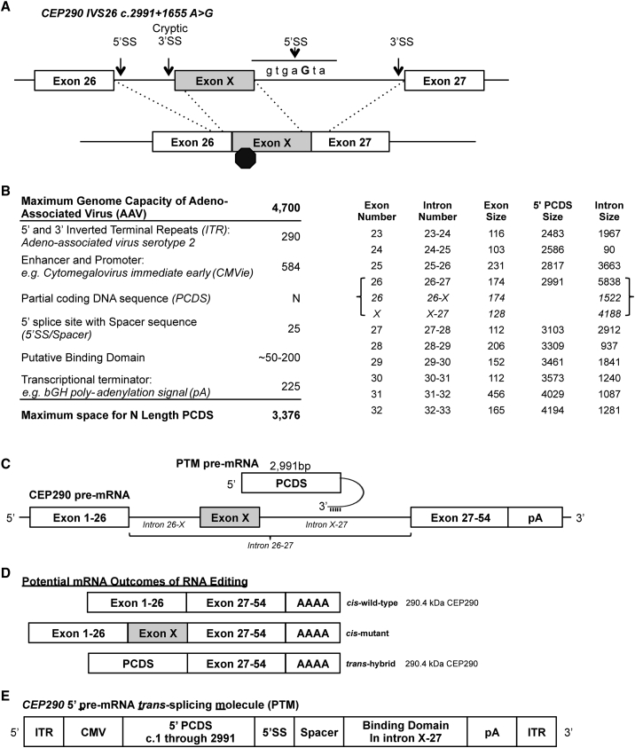
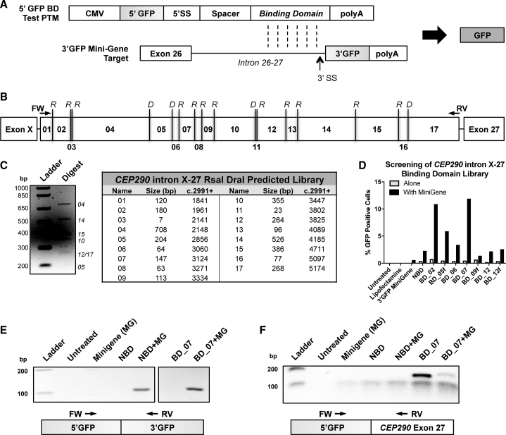
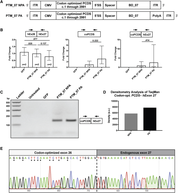
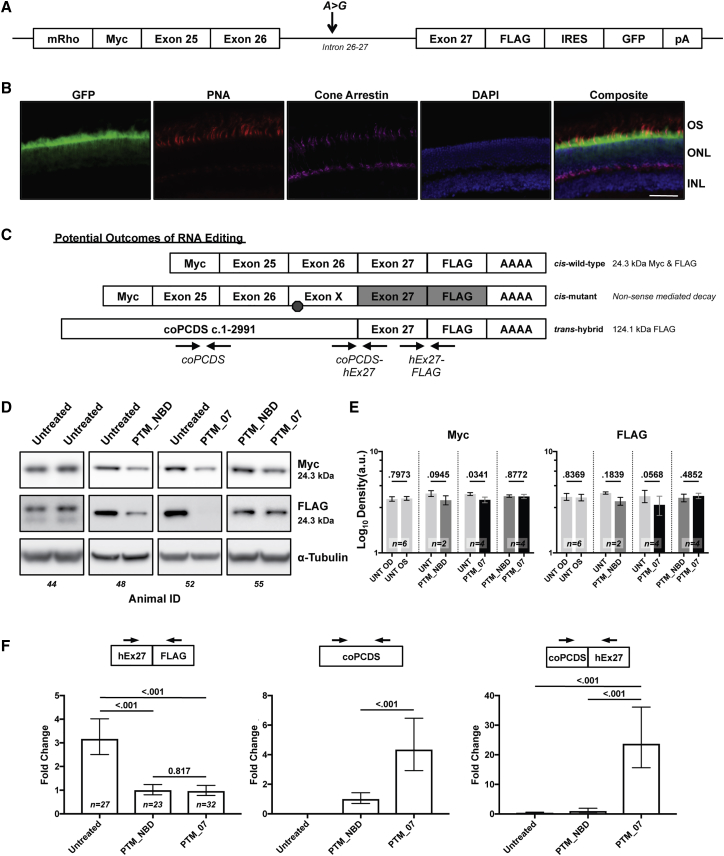
Ocular gene therapy with recombinant adeno-associated virus (AAV) has shown vector-mediated gene augmentation to be safe and efficacious in the retina in one set of diseases (retinitis pigmentosa and Leber congenital amaurosis (LCA) caused by RPE65 deficiency), with excellent safety profiles to date and potential for efficacy in several additional diseases. However, size constraints imposed by the packaging capacity of the AAV genome restrict application to diseases with coding sequence lengths that are less than 5,000 nt. The most prevalent retinal diseases with monogenic inheritance are caused by mutations in genes that exceed this capacity. Here, we designed a spliceosome mediated pre-mRNA trans-splicing strategy to rescue expression of CEP290, which is associated with Leber congenital amaurosis type 10 (LCA10) and several syndromic diseases including Joubert syndrome. We used this reagent to demonstrate editing of CEP290 in cell lines in vitro and in vivo in a mini-gene mouse model. This study is the first to show broad editing of CEP290 transcripts and in vivo proof of concept for editing of CEP290 transcripts in photoreceptors and paves the way for future studies evaluating therapeutic effects.

Keywords: CEP290; LCA10; RNA editing; animal models; cell models; gene therapy; molecular genetics; trans-splicing.


Figures

Figure 1
Figure 1
Strategy to Rescue CEP290 through 5′ Pre-mRNA trans-Splicing (A) Diagram of the most prevalent mutation in Leber congenital amaurosis type 10. This intron variant sequence (IVS) is an A-to-G transition that creates a canonical 5′ splice site (5′ SS). The novel splice site leads to inclusion of a cryptic exon, exon X. This cryptic exon encodes a premature stop codon leading to a truncated protein. (B) Left, computational analysis of required elements for a trans-splicing molecule and the size limitations of adeno-association virus. Right, analysis of CEP290 exon and intron boundaries reveals the size of a 5′ PCDS when using a 5′ trans-splicing approach to target various introns. When exon X is included in the transcript, intron 26-27 is split into intron 26-X and intron X-27. A complete table for CEP290 is included in Table 1. (C) Schematic of an approach to utilize a 5′ pre-mRNA trans-splicing molecule (PTM) to rescue mutations in CEP290 that are located 5′ to intron X-27. The PTM transcript is bound via Watson-Crick base pairing to the pre-mRNA of a target via a sequence known as the binding domain, located at the 3′ end of the PTM, and that is reverse complementary to the target sequence. (D) Three potential splicing outcomes with CEP290 IVS26 following introduction of a 5′ PTM: (1) joining of exon 26 to exon 27 from cis-splicing for the wild-type junction; (2) inclusion of exon X from cis-splicing (predominant mRNA species with IVS26 present); (3) joining of the 5′ PCDS to exon 27 from trans-splicing. Both outcomes 1 and 3 would result in full-length CEP290 peptide. (E) Illustration of an adeno-associated virus genome encompassing the therapeutically relevant CEP290 5′ trans-splicing molecule. Inverted terminal repeats (ITR) flank an expression cassette consisting of the cytomegalovirus promoter (CMV), a PCDS encoding the first 2,991 nt of the coding DNA sequence, a 5′ splice site (5′ SS), a spacer region, a binding domain that is reverse complementary to a region of intron X-27, and a polyadenylation signal sequence (pA).
Figure 2
Figure 2
Identification of a Candidate Binding Domain to Target trans-Splicing Molecules to CEP290 Intron X-27 (A) Schematic of trans-splicing between a 5′ binding domain (BD) test PTM encoding the 5′ portion of GFP (5′ GFP) and a mini-gene target encoding the 3′ portion of GFP (3′ GFP). Watson-Crick base pairing is indicated by vertical dashed lines. Trans-splicing between the two pre-mRNAs results in reconstitution and expression of GFP. (B) Diagram of RsaI (R) and DraI (D) restriction sites within a region of CEP290 intron X-27. (C) Agarose gel electrophoresis after restriction enzyme digestion of a PCR fragment corresponding to the region described in (B). The fragment was amplified from genomic DNA, digested with restriction enzymes RsaI and DraI, and visualized on a 2% TBE-agarose gel. The fragment library numbers of visible bands of expected sizes are indicated according to the table of predicted fragments. (D) Quantitation by flow cytometry of GFP expression in HEK293T transiently transfected with plasmids encoding a fragment library test PTM (gray bars) or co-transfected with a test PTM and the 3′ GFP target (black bars). Samples with “f’ demark forward orientation that is not predicted to confer trans-splicing specificity; however, BD_05f did yield a slight improvement to GFP expression over no binding domain (NBD). (E) Agarose gel electrophoresis following RT-PCR using cDNA generated from HEK293T cells transfected with plasmids in (D). Primers were designed to specifically bind to the 5′ or 3′ portions of the GFP coding DNA sequence to validate trans-splicing between the 5′ test PTM and the 3′ GFP target pre-mRNAs. Data for the other test PTMs has been removed at the break indicated. Interestingly, untargeted PTM (NBD) also resulted in trans-splicing of RNA in agreement with observed GFP expression. MG, mini-gene. (F) Samples from (E) with primers designed to specifically bind to the 5′ portion of GFP or to exon 27 of Homo sapiens CEP290. Image has been contrast enhanced to visualize the faint band in lane 7 for co-transfection of BD_07 with target mini-gene (MG).
Figure 3
Figure 3
Editing of CEP290 Transcripts Occurs in HEK293T after Transfection with PTMs (A) Illustration of adeno-associated virus genome arrangement of a CEP290 5′ PTM with binding domain 07 (PTM_07) with either no poly-adenylation signal (NPA) or with a bovine growth hormone poly-adenylation signal (PA). ITR, inverted terminal repeat; CMV, cytomegalovirus promoter; PCDS, partial coding DNA sequence of CEP290; 5′ SS, 5′ splice site. (B) qPCR was performed on HEK293T transfected with a plasmid encoding GFP as a transfection control or the plasmids in (A). TaqMan probes were designed to the junctions indicated: CEP290 exons 26 and 27 (hEx26-hEx27), a region within the 5′ codon optimized partial coding DNA sequence (coPCDS), or the novel junction of 5′ coPCDS and endogenous Homo sapiens exon 27 to signify trans-splicing (coPCDS-hEx27). Significant variation within replicates for coPCDS-hEx27 was present because the Ct was crossed at 35 cycles with either PTM; however, no amplification was observed through 40 cycles in GFP-treated samples. Samples were standardized to β-2-microglobin and normalized to NPA. Error bars are relative quantity minimum and maximum 95% confidence intervals. (C) Agarose gel electrophoresis of one of the replicate reactions from (B). (D) Densitometry analysis of the bands in (C). (E) Sanger sequencing following TOPO-cloning of the PCR product comprising the coPCDS-hEx27 junction visualized in (C). Nucleotide differences between Homo sapiens and codon-optimized CEP290 are noted by asterisks. The junction between the 5′ coPCDS and endogenous exon 27 is marked by a vertical dashed line.
Figure 4
Figure 4
HA-Tagged PTMs Show Significant Maturation following Transfection in HEK293T (A) In silico analysis of both genomes encoding either the 5′ PTM with NBD (PTM_NBD) or with CEP290 intron X-27 binding domain 07 (PTM_07) indicates an open reading frame (ORF) is present that is predicted to encode a 118.7-kDa peptide if the pre-mRNA matures without trans-splicing. (B) Representative western blot images of HA-tag and CEP290 using extracts from HEK293T harvested 72 hr following transfection with the indicated plasmids. HA-tagged CEP290 resulting from trans-splicing was expected at ∼290 kDa. α-tubulin was used as a loading control. (C) Densitometry quantification of CEP290 and the 118.7-kDa bands. Values per replicate group were standardized to α-tubulin and normalized to PTM_NBD. Error bars are SD; n = 6 independent experiments. p < 0.05 was considered significant.
Figure 5
Figure 5
Editing of Mini-CEP290 Transcripts Occurs In Vivo following Sub-retinal Injection of 7m8AAV-5′ PTMs (A) Diagram of the CEP290 intron 26 mini-gene. The mini-gene is driven by the murine Rhodopsin promoter (mRho). CEP290 exons 25 and 26 are joined and followed by the complete intron 26-27 and exon 27. An A > G mutation corresponding to c.2991+1655 was included to assess exon X splicing in the model. An amino-terminal Myc and a carboxy-terminal FLAG tag were added to flank the mini-gene. Additionally, an internal ribosomal entry site (IRES) was added to drive expression of EGFP and is terminated by a bovine growth hormone poly-adenylation signal sequence (pA). (B) Immunofluorescence staining of a retinal cross-section from a mini-CEP290 mouse. OS, outer segment. ONL, outer nuclear layer. INL, inner nuclear layer. Scale bar is 50 μm. (C) Illustration of potential splicing outcomes within the mini-CEP290 mouse. Canonical cis-splicing would result in a predicted 24.3-kDa peptide. Alternative cis-splicing with exon X would yield a predicted non-sense media decay transcript encoding a truncated peptide with Myc at 17.1 kDa and no FLAG translation. Trans-splicing with a 5′ PTM would replace the amino-terminal Myc and generate a 124.1-kDa peptide with a FLAG tag. Arrow pairs indicate locations of PCR primers used to detect specific splicing events. (D) Representative western blot images of Myc and FLAG at 24.3 kDa showing OD and OS lanes per animal for each contralateral treatment cohort. (E) Densitometry quantification showing the mean log10 values of each contralateral treatment cohort. Samples were standardized to α-tubulin. Error bars are SEM. Sample sizes as indicated. Individual animal data is available in Figure S1A. (F) qPCR from cDNA generated by RNA extracts from whole eyes of mini-CEP290 mice. TaqMan probes were designed to the junctions of Homo sapiens exon 27 and FLAG to detect total expression of the mini-gene (left) to a region within the PCDS to detect total expression of the PTM (center) or to the novel junction of codon-optimized CEP290 PCDS and Homo sapiens CEP290 exon 27 (right). Values from treatment-matched samples were averaged as biological groups, standardized to murine β-2-microglobin and normalized to PTM_NBD. p < 0.05 was considered significant. Error bars are relative quantity minimum and maximum 95% confidence intervals. Sample sizes as indicated. Individual animal data is available in Figure S1B.

Similar articles

Cited by

References

    1. Koenekoop R.K. An overview of Leber congenital amaurosis: a model to understand human retinal development. Surv. Ophthalmol. 2004;49:379–398. - PubMed
    1. Simonelli F., Maguire A.M., Testa F., Pierce E.A., Mingozzi F., Bennicelli J.L., Rossi S., Marshall K., Banfi S., Surace E.M. Gene therapy for Leber’s congenital amaurosis is safe and effective through 1.5 years after vector administration. Mol. Ther. 2010;18:643–650. - PMC - PubMed
    1. Bennett J., Wellman J., Marshall K.A., McCague S., Ashtari M., DiStefano-Pappas J., Elci O.U., Chung D.C., Sun J., Wright J.F. Safety and durability of effect of contralateral-eye administration of AAV2 gene therapy in patients with childhood-onset blindness caused by RPE65 mutations: a follow-on phase 1 trial. Lancet. 2016;388:661–672. - PMC - PubMed
    1. Russell S., Bennett J., Wellman J.A., Chung D.C., Yu Z.F., Tillman A., Wittes J., Pappas J., Elci O., McCague S. Efficacy and safety of voretigene neparvovec (AAV2-hRPE65v2) in patients with RPE65-mediated inherited retinal dystrophy: a randomised, controlled, open-label, phase 3 trial. Lancet. 2017;390:849–860. - PMC - PubMed
    1. den Hollander A.I., Black A., Bennett J., Cremers F.P.M. Lighting a candle in the dark: advances in genetics and gene therapy of recessive retinal dystrophies. J. Clin. Invest. 2010;120:3042–3053. - PMC - PubMed