Review
. 2018 Nov;84(5):602-610.
doi: 10.1038/s41390-018-0141-6. Epub 2018 Sep 8.

Neonatal cerebrovascular autoregulation

Affiliations
Review

Neonatal cerebrovascular autoregulation

Christopher J Rhee et al. Pediatr Res. 2018 Nov.

Abstract

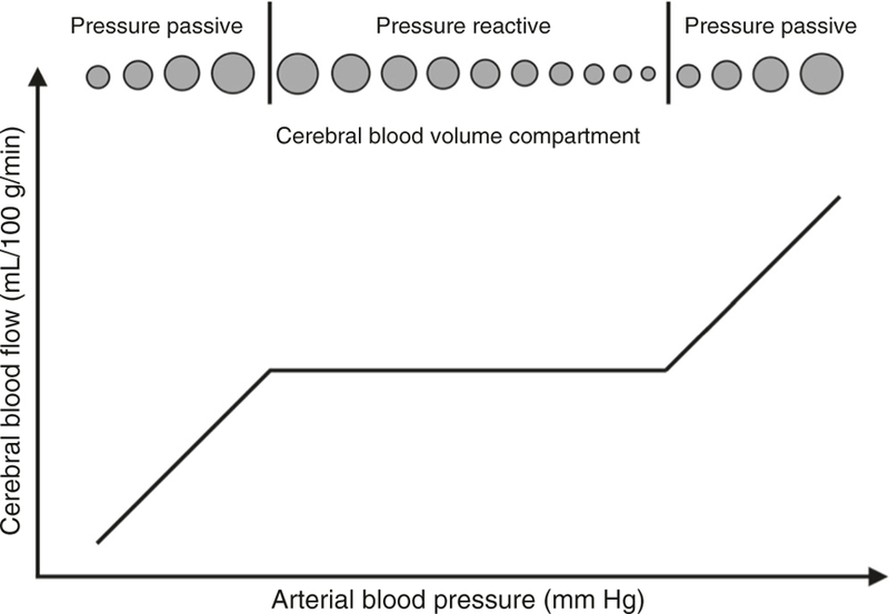
Cerebrovascular pressure autoregulation is the physiologic mechanism that holds cerebral blood flow (CBF) relatively constant across changes in cerebral perfusion pressure (CPP). Cerebral vasoreactivity refers to the vasoconstriction and vasodilation that occur during fluctuations in arterial blood pressure (ABP) to maintain autoregulation. These are vital protective mechanisms of the brain. Impairments in pressure autoregulation increase the risk of brain injury and persistent neurologic disability. Autoregulation may be impaired during various neonatal disease states including prematurity, hypoxic-ischemic encephalopathy (HIE), intraventricular hemorrhage, congenital cardiac disease, and infants requiring extracorporeal membrane oxygenation (ECMO). Because infants are exquisitely sensitive to changes in cerebral blood flow (CBF), both hypoperfusion and hyperperfusion can cause significant neurologic injury. We will review neonatal pressure autoregulation and autoregulation monitoring techniques with a focus on brain protection. Current clinical therapies have failed to fully prevent permanent brain injuries in neonates. Adjuvant treatments that support and optimize autoregulation may improve neurologic outcomes.


Figures

Fig. 1
Fig. 1
Cerebral blood flow autoregulation curve. Autoregulation is the physiologic mechanism that holds the blood flow to the brain constant across a range of blood pressure. The flat area of the curve is a state of pressure reactivity where blood pressure and blood flow go in opposite directions. However, outside the range of auto-regulation on the two ends is a state of pressure passivity where blood flow is purely dependent on blood pressure. There is no more reactivity in the vessels
Fig. 2
Fig. 2
Changes in critical closing pressure across gestational age. Critical closing pressure increases with gestational age at a rate of 1.4 mmHg per week of gestation (P < 0.0001). From Rhee et al.
Fig. 3
Fig. 3
Graphical explanations of the autoregulation metrics. a Coherence analysis of CBF measured by transcranial Doppler (TCD) ultrasound in preterm neonate. Coherence is high between ABP (red line) and CBF (black line), indicating a state of dysautoregulation at slow wave frequencies. Bottom panel shows coherence as a function of frequency. b Waveform 1 (W1, solid line) and waveform 2 (W2, dotted line) are shown for concept. The gain is the damping effect and the phase shift is the time delay between the two waveforms. Lower gain indicates better autoregulatory function. c, d Blood pressure was decreased in a neonatal piglet with continuous mean arterial blood pressure (MAP), intracranial pressure (ICP), cerebral blood volume (CBV) NIRS tissue hemoglobin index, and cerebral blood flow (CBF) laser Doppler flow (LDF) measurements. When MAP exceeds the lower limit of autoregulation and CBF is relatively constant, slow MAP waves are out of phase with the ICP and CBV (red star and box). As MAP decreases below the lower limit of autoregulation and CBF falls, the MAP, ICP, and CBV waves are all in phase, indicating a state of dysautoregulation (blue cross and box). Adapted with permission from: SAGE Publications et al, American Physiological Society et al,, and Wolters Kuwer et al
Fig. 4
Fig. 4
Determining optimal arterial blood pressure. An example of one patient using autoregulation monitoring to determine optimal mean arterial blood pressure (MAP). Slow waves of MAP (top) and cerebral blood volume (CBV, bottom) are depicted (a–c). Autoregulation monitoring during periods of intact and impaired autoregulation in this infant. When autoregulation is intact, MAP waves negatively correlate to changes in NIRS-measured CBV (a, b). The correlation of CBV and MAP is the hemoglobin volume index (HVx). When autoregulation is impaired, MAP and CBV become positively correlated, yielding a positive HVx (c, d). Thus, a more negative HVx indicates functional pressure reactivity and intact autoregulation, and a more positive HVx indicates decreased pressure reactivity and impaired autoregulation. When the measures of HVx are plotted as a function of MAP and placed in 5-mmHg bins, optimal MAP is identified as the nadir when the correlation coefficients are plotted against ABP (e). Here for this patient, optimal ABP is 50 mmHg, where autoregulation is intact and most robust

References

    1. Helou S, Koehler RC, Gleason CA, Jones MD & Traystman RJ Cerebrovascular autoregulation during fetal development in sheep. Am. J. Physiol. Heart C 266, H1069–H1074 (1994). - PubMed
    1. Rhee CJ et al. The ontogeny of cerebrovascular pressure autoregulation in human premature infants. J. Perinatol 34, 926–931 (2014). - PMC - PubMed
    1. Rorke LB Anatomical features of the developing brain implicated in pathogenesis of hypoxic-ischemic injury. Brain. Pathol 2, 211–221 (1992). - PubMed
    1. Wladimiroff JW & van Bel F Fetal and neonatal cerebral blood flow. Semin. Perinatol 11, 335–346 (1987). - PubMed
    1. Pryds O & Edwards AD Cerebral blood flow in the newborn infant. Arch. Dis. Child. Fetal Neonatal Ed 74, F63–F69 (1996). - PMC - PubMed

Publication types

LinkOut - more resources