. 2018 Nov 6;26(11):1499-1512.e5.
doi: 10.1016/j.str.2018.07.012. Epub 2018 Sep 6.

Trypanosomatid Deoxyhypusine Synthase Activity Is Dependent on Shared Active-Site Complementation between Pseudoenzyme Paralogs

Affiliations

Trypanosomatid Deoxyhypusine Synthase Activity Is Dependent on Shared Active-Site Complementation between Pseudoenzyme Paralogs

Gustavo A Afanador et al. Structure. .

Abstract

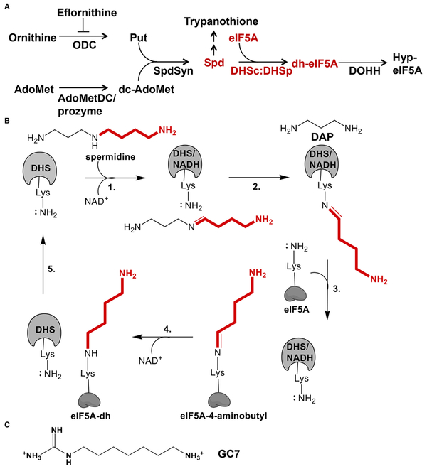
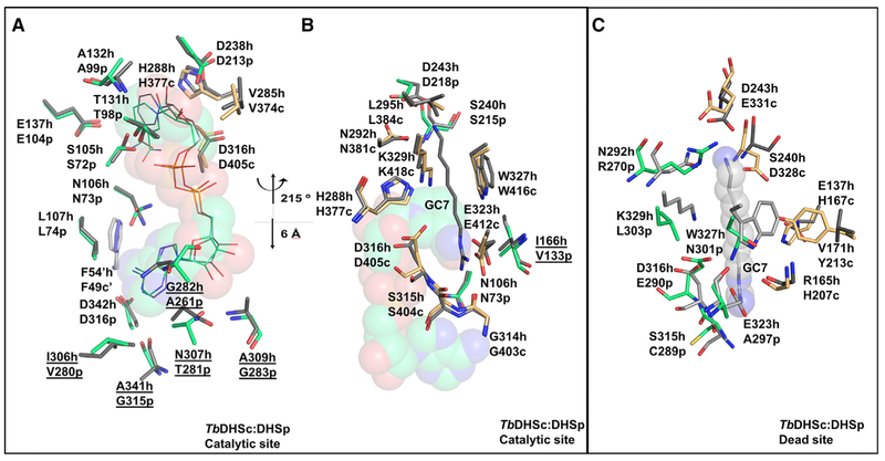
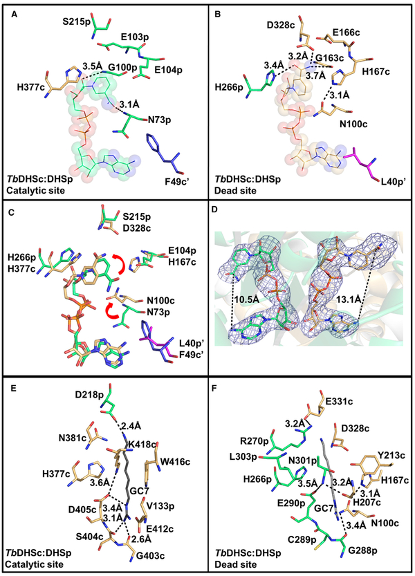
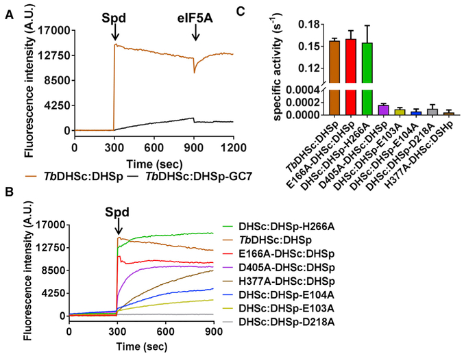
Trypanosoma brucei is a neglected tropical disease endemic to Africa. The polyamine spermidine is essential for post-translational hypusine modification of eukaryotic initiation factor 5A (eIF5A), which is catalyzed by deoxyhypusine synthase (TbDHS). In trypanosomatids, deoxyhypusine synthase (DHS) activity is dependent on heterotetramer formation between two paralogs, DHSc and DHSp, both with minimal activity on their own due to missing catalytic residues. We determined the X-ray structure of TbDHS showing a single functional shared active site is formed at the DHSc/DHSp heterodimer interface, with deficiencies in one subunit complemented by the other. Each heterodimer contains two NAD+ binding sites, one housed in the functional catalytic site and the second bound in a remnant dead site that lacks key catalytic residues. Functional analysis of these sites by site-directed mutagenesis identified long-range contributions to the catalytic site from the dead site. Differences between trypanosomatid and human DHS that could be exploited for drug discovery were identified.

Keywords: Trypanosoma brucei; deoxyhypusine synthase; eIF5A; hypusine; polyamines; pseudoenzyme; trypanosomatids.


Figures

Figure 1.
Figure 1.. Polyamine Pathway and Reaction Mechanism of DHS
(A) Polyamine and hypusine biosynthetic pathways in trypanosomatids. Ornithine decarboxylase (ODC), S-adenosylmethionine decarboxylase (AdoMetDC), composed of a heterodimer of AdoMetDCc and AdoMetDCp, spermidine synthase (SpdSyn), eukaryotic initiation factor 5A (eIF5A), deoxyhypusine synthase (DHS), and deoxyhypusine hydroxylase (DOHH). (B) DHS catalyzed reaction mechanism. AdoMet, S-adenosylmethionine; Put, putrescine; Spd, spermidine; DAP, diaminopropane. (C) Chemical structure of GC7, DHS inhibitor, and non-hydrolysable spermidine analog.
Figure 2.
Figure 2.. Structure-Based Sequence Alignment of hDHS (PDB: 1DHS), TbDHSc, and TbDHSp
Residues highlighted show the NAD+ binding site (yellow), spermidine/GC7 binding site (cyan), and both ligands (green) from hDHS (PDB: 1RQD). The catalytic Lys is identified with the symbol ¥. Residues targeted for TbDHSc:DHSp mutagenesis are indicated with arrows. Helices are labeled with “H” and α sheets with “B.” B1* and B3* are found only in DHSc, H1’ is only in hDHS, and H6Λ^ is in both hDHS and DHSp. Structural alignment by PROMALS3D (Pei et al., 2008). Residues not resolved in the structure are underlined.
Figure 3.
Figure 3.. Tetrameric and Monomeric Configurations of TbDHSc:DHSp
(A) Cartoon representation of hDHSand TbDHSc:DHSp. hDHS (gray)forms two equivalent functional active sites (catalytic site) in each of the two dimer interfaces of the homotetramer. For TbDHSciDHSp one catalytic site and one dead site are formed between DHSc (orange) and DHSp (green) in the dimer interface; the tetramer is composed of a dimer of heterodimers. (B)Heterotetrameric structure of TbDHSc:DHSp as a ribbon diagram with the four NAD+ shown as spheres. Helices found only in DHSc are labeled H7 and H8. (C) Alignment of TbDHSc and TbDHSp monomers: 1,575 atoms aligned, RMSD of 1.1 Å. (D) Alignment of hDHS (PDB: 1DHS) and TbDHSc monomers: 1,795 atoms aligned, RMSD of 0.8 Å. (E) Alignment of hDHS (PDB: 1DHS) and TbDHSp monomers: 1,804 atoms aligned, RMSD of 0.8 Å. See also Figure S1.
figure 4.
figure 4.. TbDHSc:DHSp Contains One Catalytic Site and One Dead Site at the Dimer Interface
(A) Heterodimer of TbDHSc (orange) and TbDHSp (green) (ribbon diagram) with two NAD+ (spheres). (B) Dimer cartoon showing NAD+ sites and the putative spermidine binding site (based on the hDHS structure). Amino acid residues targeted for site-directed mutagenesis (Table 2) are shown. Residues conserved in both subunits are marked by the same symbol (# or Λ). The catalytic Lys (K418) and the corresponding residue in the dead site (L303) are labeled with an asterisk (*) and underlined. (C) NAD+ binding sites at the dimer interface of TbDHSc:DHSp. Two residues (marked by ‘) that are contributed from the opposite dimer are also shown (F49-DHSc [blue] and DHSp-L40 [magenta]).
Figure 5.
Figure 5.. Comparison of the TbDHSc:DHSp and hDHS
(A and B) Comparison of TbDHSc:DHSp and hDHS catalytic sites. (A) NAD+ binding site, in the same orientation as the catalytic site in Figure 4C. TbDHSc:DHSp and hDHS (PDB: 1DHS) tetramers were superimposed in PyMol using only TbDHSp with 283 atoms aligned to an RMSD = 0.74 Å. Amino acid differences between hDHS and TbDHSare underlined. (B) GC7 binding site. This view is rotated around the vertical axis by approximately 215° and translated approximately 6 Å from (A), as indicated. TbDHSc:DHSp and hDHS (PDB: 1RQD) were superimposed as above to RMSD = 0.8 Å. hDHS is in gray, TbDHSc is in orange, and TbDHSp is in green. GC7 is displayed as gray sticks. The NAD+ is displayed as spheres. (C) Comparison of the TbDHSc:DHSp dead site with the hDHS catalytic site. The hDHS spermidine binding pocket was aligned with the TbDHSc:DHSp dead-site pocket. Select residues within the 4-Å shell of bound ligands are shown.
Figure 6.
Figure 6.. Comparison of NAD+ and GC7 Binding Sites between TbDHSc and TbDHSp Sites
(A) NAD+ binding pocket in the catalytic site. (B) NAD+ binding pocket in the dead site. NAD+ (spheres and lines), TbDHSc (orange sticks), and TbDHSp (green sticks). F49-DHSc (blue) and DHSp-L40 (magenta) are from the opposite dimer. (C) Overlay of the catalytic and dead sites highlighting the nicotinamide flip (red arrows). (D) Polder omit map (blue mesh) for NAD (Liebschner et al., 2017) contoured at 4× the RMS level. The map was calculated in the program suite Phenix (Adams et al., 2010) by omitting the coordinates for NAD+. (E) GC7 binding pocket in the catalytic site. GC7 (gray sticks) is from hDHS (PDB: 1RQD) based on the superimposition described in Figure 5B. (F) Dead-site amino acids in the region of the GC7 site showing the highly variable amino acid composition relative to the catalytic site. Orientation is similar to Figure 5B and (E).
Figure 7.
Figure 7.. Single-Turnover and Steady-State Kinetic Analysis of TbDHSc:DHSp WT and Mutant Enzymes
(A) Single-turnover NADH formation for WT TbDHSc:DHSp. NADH formation was monitored by fluorescence under single-turnover conditions for excitation at 350 nm and emission at 441 nm. Spermidine (Spd) (1 mM) was added to enzyme (1 μM) and NAD+ (1 mM) or eIF5A (15 μM) at the indicated time (orange line). Pre-incubation of TbDHSc:DHSp with 10 μM GC7 inhibits the reaction (black line). Experiments were done in duplicate and traces represent the average of the duplicate. (B) Single-turnover NADH formation for TbDHSc:DHSp mutant enzymes. Mutant enzyme pairs are identified with symbols (Λ and #) as shown in Figure 4C. (C) Steady-state kinetic analysis of WT and mutant DHS activity. For WT and active mutants, [DHS] = 5 nM, while inactive mutants were assayed at 1 μM. Substrate concentrations were 1 mM NAD+, 78 μM spermidine, and 15 μM eIF5A (KM = 0.7 μM), which represent saturating concentrations for the WT enzyme (Nguyen et al., 2013). Data were collected in triplicate, and error bars represent SEM. See also Tables S1 and S2.

Comment in

References

    1. Adams PD, Afonine PV, Bunkoczi G, Chen VB, Davis IW, Echols N, Headd JJ, Hung LW, Kapral GJ, Grosse-Kunstleve RW, et al. (2010). PHENIX: a comprehensive Python-based system for macromolecular struc-ture solution. Acta Crystallogr. D Biol. Crystallogr 66, 213–221. - PMC - PubMed
    1. Afanador GA., Muench SP., McPhillie M., Fomovska A., Schon A., Zhou Y., Cheng G., Stec J., Freundlich JS., Shieh HM., et al. (2013). Discrimination of potent inhibitors of Toxoplasma gondii enoyl-acyl carrier protein reductase by a thermal shift assay. Biochemistry 52, 9155–9166. - PMC - PubMed
    1. Borek D, Cymborowski M, Machius M, Minor W, and Otwinowski Z (2010). Diffraction data analysis in the presence of radiation damage. Acta Crystallogr. D Biol. Crystallogr 66, 426–36. - PMC - PubMed
    1. Borek D, Dauter Z, and Otwinowski Z (2013). Identification of patterns in diffraction intensities affected by radiation exposure. J. Synchrotron Radiat 20, 37–8. - PMC - PubMed
    1. Borek D, Minor W, and Otwinowski Z (2003). Measurement errors and their consequences in protein crystallography. Acta Crystallogr. D Biol. Crystallogr 59,2031–2038. - PubMed

Publication types

MeSH terms

LinkOut - more resources