

Rapid Mast Cell Generation from Gata2 Reporter Pluripotent Stem Cells
- PMID: 30197119
- PMCID: PMC6178197
- DOI: 10.1016/j.stemcr.2018.08.007
Rapid Mast Cell Generation from Gata2 Reporter Pluripotent Stem Cells
Abstract
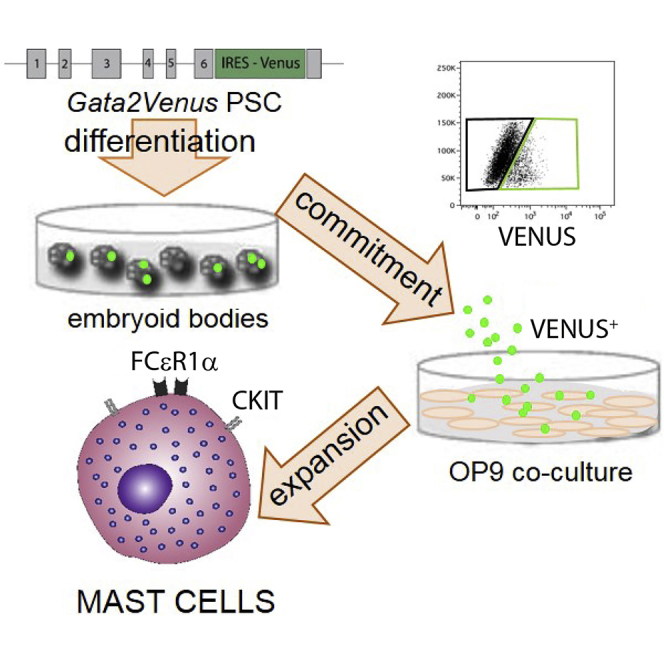
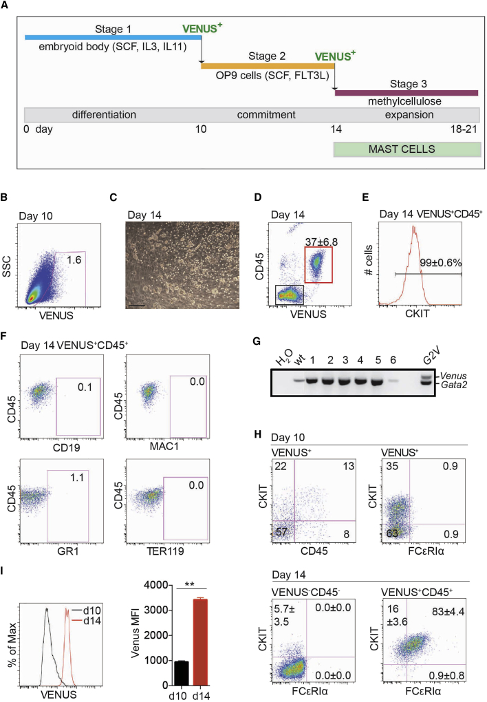
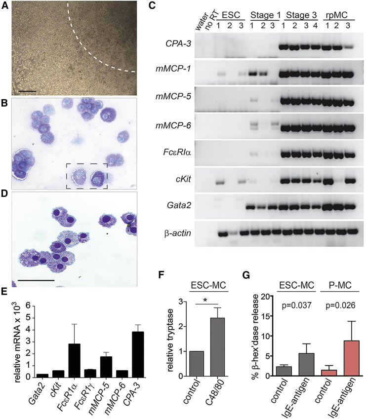
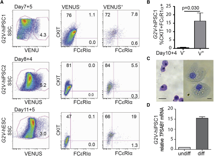
Mast cells are tissue-resident immune cells. Their overgrowth/overactivation results in a range of common distressing, sometimes life-threatening disorders, including asthma, psoriasis, anaphylaxis, and mastocytosis. Currently, drug discovery is hampered by use of cancer-derived mast cell lines or primary cells. Cell lines provide low numbers of mature mast cells and are not representative of in vivo mast cells. Mast cell generation from blood/bone marrow gives poor reproducibility, requiring 8-12 weeks of culture. Here we report a method for the rapid/robust production of mast cells from pluripotent stem cells (PSCs). An advantageous Gata2Venus reporter enriches mast cells and progenitors as they differentiate from PSCs. Highly proliferative mouse mast cells and progenitors emerge after 2 weeks. This method is applicable for rapid human mast cell generation, and could enable the production of sufficient numbers of physiologically relevant human mast cells from patient induced PSCs for the study of mast cell-associated disorders and drug discovery.
Keywords: ESC; Gata2; Venus reporter; differentiation; iPSC; immune effectors; innate immune cells; mast cells; rapid protocol; stem cells.
Copyright © 2018 The Authors. Published by Elsevier Inc. All rights reserved.
Figures
Similar articles
-
In Vitro Differentiation of Gata2 and Ly6a Reporter Embryonic Stem Cells Corresponds to In Vivo Waves of Hematopoietic Cell Generation.Stem Cell Reports. 2018 Jan 9;10(1):151-165. doi: 10.1016/j.stemcr.2017.11.018. Epub 2017 Dec 21. Stem Cell Reports. 2018. PMID: 29276152 Free PMC article.
-
The STAT5-GATA2 pathway is critical in basophil and mast cell differentiation and maintenance.J Immunol. 2015 May 1;194(9):4328-38. doi: 10.4049/jimmunol.1500018. Epub 2015 Mar 23. J Immunol. 2015. PMID: 25801432 Free PMC article.
-
GATA2 is critical for the maintenance of cellular identity in differentiated mast cells derived from mouse bone marrow.Blood. 2015 May 21;125(21):3306-15. doi: 10.1182/blood-2014-11-612465. Epub 2015 Apr 8. Blood. 2015. PMID: 25855601
-
Cytokines regulate development of human mast cells from hematopoietic progenitors.Int J Hematol. 2002 May;75(4):350-6. doi: 10.1007/BF02982123. Int J Hematol. 2002. PMID: 12041663 Review.
-
Differentiation of pluripotent stem cells into endothelial cells.Curr Opin Hematol. 2015 May;22(3):252-7. doi: 10.1097/MOH.0000000000000140. Curr Opin Hematol. 2015. PMID: 25767955 Free PMC article. Review.
Cited by
-
Understanding and Modulating Immunity With Cell Reprogramming.Front Immunol. 2019 Dec 11;10:2809. doi: 10.3389/fimmu.2019.02809. eCollection 2019. Front Immunol. 2019. PMID: 31921109 Free PMC article. Review.
-
A Novel GATA2 Protein Reporter Mouse Reveals Hematopoietic Progenitor Cell Types.Stem Cell Reports. 2020 Aug 11;15(2):326-339. doi: 10.1016/j.stemcr.2020.06.008. Epub 2020 Jul 9. Stem Cell Reports. 2020. PMID: 32649900 Free PMC article.
-
Neuroinflammation in Amyotrophic Lateral Sclerosis and Frontotemporal Dementia and the Interest of Induced Pluripotent Stem Cells to Study Immune Cells Interactions With Neurons.Front Mol Neurosci. 2021 Dec 14;14:767041. doi: 10.3389/fnmol.2021.767041. eCollection 2021. Front Mol Neurosci. 2021. PMID: 34970118 Free PMC article. Review.
-
Generating hematopoietic cells from human pluripotent stem cells: approaches, progress and challenges.Cell Regen. 2023 Sep 1;12(1):31. doi: 10.1186/s13619-023-00175-6. Cell Regen. 2023. PMID: 37656237 Free PMC article. Review.
-
Iterative Single-Cell Analyses Define the Transcriptome of the First Functional Hematopoietic Stem Cells.Cell Rep. 2020 May 12;31(6):107627. doi: 10.1016/j.celrep.2020.107627. Cell Rep. 2020. PMID: 32402290 Free PMC article.
References
-
- Akashi K., Traver D., Miyamoto T., Weissman I.L. A clonogenic common myeloid progenitor that gives rise to all myeloid lineages. Nature. 2000;404:193–197. - PubMed
-
- Bischoff S.C. Role of mast cells in allergic and non-allergic immune responses: comparison of human and murine data. Nat. Rev. Immunol. 2007;7:93–104. - PubMed
Publication types
MeSH terms
Substances
Grants and funding
LinkOut - more resources
Full Text Sources
Other Literature Sources
