. 2018 Oct 2;115(7):1231-1239.
doi: 10.1016/j.bpj.2018.07.039. Epub 2018 Aug 17.

Human Dystrophin Structural Changes upon Binding to Anionic Membrane Lipids

Affiliations

Human Dystrophin Structural Changes upon Binding to Anionic Membrane Lipids

Raphael Dos Santos Morais et al. Biophys J. .

Abstract

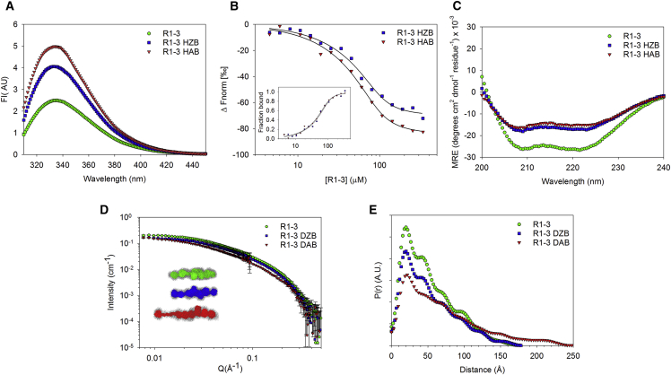
Scaffolding proteins play important roles in supporting the plasma membrane (sarcolemma) of muscle cells. Among them, dystrophin strengthens the sarcolemma through protein-lipid interactions, and its absence due to gene mutations leads to the severe Duchenne muscular dystrophy. Most of the dystrophin protein consists of a central domain made of 24 spectrin-like coiled-coil repeats (R). Using small angle neutron scattering (SANS) and the contrast variation technique, we specifically probed the structure of the three first consecutive repeats 1-3 (R1-3), a part of dystrophin known to physiologically interact with membrane lipids. R1-3 free in solution was compared to its structure adopted in the presence of phospholipid-based bicelles. SANS data for the protein/lipid complexes were obtained with contrast-matched bicelles under various phospholipid compositions to probe the role of electrostatic interactions. When bound to anionic bicelles, large modifications of the protein three-dimensional structure were detected, as revealed by a significant increase of the protein gyration radius from 42 ± 1 to 60 ± 4 Å. R1-3/anionic bicelle complexes were further analyzed by coarse-grained molecular dynamics simulations. From these studies, we report an all-atom model of R1-3 that highlights the opening of the R1 coiled-coil repeat when bound to the membrane lipids. This model is totally in agreement with SANS and click chemistry/mass spectrometry data. We conclude that the sarcolemma membrane anchoring that occurs during the contraction/elongation process of muscles could be ensured by this coiled-coil opening. Therefore, understanding these structural changes may help in the design of rationalized shortened dystrophins for gene therapy. Finally, our strategy opens up new possibilities for structure determination of peripheral and integral membrane proteins not compatible with different high-resolution structural methods.


Figures

Figure 1
Figure 1
(A) Schematic representation of dystrophin and its four domains, including the central domain composed of 24 spectrin-like repeats (R). The R1–3 protein fragment is framed in red and can interact with membrane phospholipids. LBD, lipid binding domain; ABD, actin binding domain. (B) The 3D structure of three spectrin repeats folded in triple coiled-coil (PDB: 1U4Q) showing the organization of dystrophin central domain is shown. The linker region is the junction between the helix C of one repeat and the helix A of the subsequent one. To see this figure in color, go online.
Figure 2
Figure 2
Characterization of the protein/lipid interactions of the R1–3 dystrophin fragment. (A) The Trp fluorescence intensity of R1–3 in the absence (green circles) or in the presence of zwitterionic (HZB, blue squares) or anionic (HAB, red triangles) bicelles is shown. (B) MST data is shown; the dissociation constant (Kd) was determined to be ∼10 μM for the protein/bicelle complex. The inset shows the same MST data but using the “fraction bound versus protein concentration” representation. (C) Far-UV CD spectra with the same legend as for fluorescence intensity are shown. CD spectra highlight that the α-helical folding of R1–3 is maintained upon lipid binding. (D) SANS intensities measured for R1–3 alone in solution (green circles) or in interaction with contrast-matched deuterated zwitterionic (DZB, blue squares) or anionic (DAB, red triangles) bicelles are shown. The inset shows ab initio shapes of (from top to bottom) R1–3 free in solution (green) or in interaction with DZB (blue) or DAB (red), corresponding to the DAMMIF models obtained with the smallest normalized spatial discrepancy and surrounded by the corresponding DAMAVER model (gray). (E) The pair-distribution function P(r) analysis with the same color code showing an increase of Dmax of R1–3 when the protein fragment is in interaction with anionic bicelles is shown. To see this figure in color, go online.
Figure 3
Figure 3
(A) Final structural model obtained by CG-MD of R1–3 bound to anionic bicelles (HAB). The protein is shown in green, with orange spots corresponding to the pacFA PC cross-linked regions identified by MS. Lipids are colored in blue for DMPC, red for DMPS, and cyan for DHPC. (B) The final structural model obtained by CG-IMD for the R1–3/anionic bicelle complex is shown; HA, HB, and HC helices of each repeat are shown in yellow, purple, and green, respectively. (C) A collection of the five protein all-atom models obtained after opening the 1HA and 1HB helical regions of the R1 repeat through CG-IMD simulations is shown. (D) Experimental SANS intensities (circles and triangles for R1–3 alone or bound to contrast-matched anionic bicelles, respectively) are shown fitted with the theoretical CRYSON curves generated from the R1–3 model free in solution (green) and the R1–3 model bound to anionic bicelles (purple). The inset shows the corresponding all-atom models represented with the same color code. Blue and red dots represent the N- and C-termini, respectively. The peptides bearing the pacFA PC are colored in orange, and the putative ALPS motif (QGRVGNILQLGSKLIGTG) is colored in red. To see this figure in color, go online.

References

    1. Seddon A.M., Curnow P., Booth P.J. Membrane proteins, lipids and detergents: not just a soap opera. Biochim. Biophys. Acta. 2004;1666:105–117. - PubMed
    1. Hendrickson W.A. Atomic-level analysis of membrane-protein structure. Nat. Struct. Mol. Biol. 2016;23:464–467. - PMC - PubMed
    1. Fernandez-Leiro R., Scheres S.H. Unravelling biological macromolecules with cryo-electron microscopy. Nature. 2016;537:339–346. - PMC - PubMed
    1. Petoukhov M.V., Svergun D.I. Analysis of X-ray and neutron scattering from biomacromolecular solutions. Curr. Opin. Struct. Biol. 2007;17:562–571. - PubMed
    1. Pérez J., Koutsioubas A. Memprot: a program to model the detergent corona around a membrane protein based on SEC-SAXS data. Acta Crystallogr. D Biol. Crystallogr. 2015;71:86–93. - PMC - PubMed

Publication types