Review
. 2018 Dec;36(12):1259-1274.
doi: 10.1016/j.tibtech.2018.07.007. Epub 2018 Sep 6.

Smart Bandages: The Future of Wound Care

Affiliations
Review

Smart Bandages: The Future of Wound Care

Hossein Derakhshandeh et al. Trends Biotechnol. 2018 Dec.

Abstract

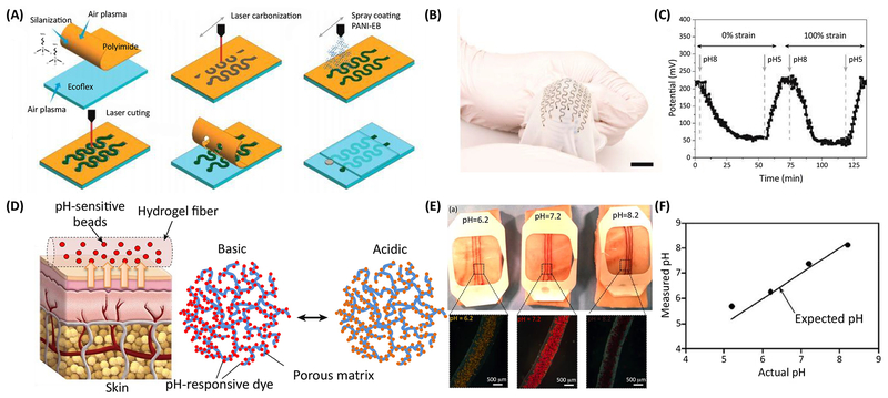
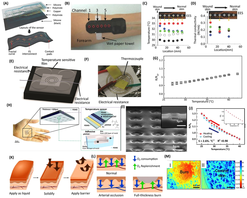
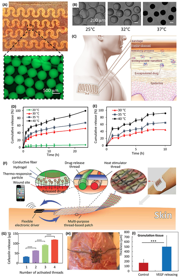
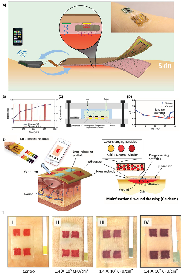
Chronic non-healing wounds are major healthcare challenges that affect a noticeable number of people; they exert a severe financial burden and are the leading cause of limb amputation. Although chronic wounds are locked in a persisting inflamed state, they are dynamic and proper therapy requires identifying abnormalities, administering proper drugs and growth factors, and modulating the conditions of the environment. In this review article, we discuss technologies that have been developed to actively monitor the wound environment. We also highlight drug delivery tools that have been integrated with bandages to facilitate precise temporal and spatial control over drug release and review automated or semi-automated systems that can respond to the wound environment.

Keywords: drug delivery; flexible electronics; integrated systems; smart bandages; wound care.


Figures

Figure 1.
Figure 1.
Examples of bandages with integrated pH sensors. (a) Schematic view of the fabrication process of highly stretchable potentiometric pH sensors. (b) Photograph of a typical bandage with integrated pH sensor placed on a curved surface demonstrating its stretchability and flexibility. (c) Real-time monitoring of the pH and the sensor response to pH changes under various mechanical strains showing negligible effect of strain on the sensor readout. (d) A colorimetric pH sensor based hydrogels carrying beads loaded with pH-sensitive dye for long-term monitoring of wound. (e) Images demonstrating color changes in of pH sensitive dressings on the pig skin when sprayed with solutions of different pH values. (f) The data extracted from the images taken using a smartphone in comparison to the actual pH values of the sprayed solutions. Figures are reproduced with permission from [39], [40].
Figure 2.
Figure 2.
Examples of wound dressings capable of monitoring skin temperature and oxygenation. (a) Schematic illustration of microfabrication of flexible temperature sensor. (b) A typical fabricated bandage that could maintain conformal contact with the skin. (c,d) The distribution of cutaneous temperature and thermal conductivity measured using the bandage. (e) Schematic illustration of a temperature sensor fabricated on an elastic nanofibrous substrate. (f) A representative image of a sensor fabricated by screen printing of silver ink on the nanofibrous substrate. (g) Changes in the relative electrical resistance of the fabricated sensor normalized to its reference resistance at 37 °C. (h) Image of the wearable temperature sensor attached on the skin and schematic illustration of the different layers of the fabricated temperature sensors. (i) A SEM image of the octopus mimicking surface applying suction for enhanced adhesion to the skin surface and forming conformal contact. (j) The variation of the relative resistance of the integrated sensor with temperature. (k) Schematic illustration of the application of the oxygen sensing of liquid-based bandage. (l) Schematic demonstrating of the tissue oxygenation in various types of wounds. (m) Oxygen consumption measured using the bandage in burn tissue (I) and healthy tissue (II). Blue depicts higher oxygen consumption and red indicates lower oxygen consumption in the tissue. Figures are reproduced with permission from [44], [42], [57], [45].
Figure 3.
Figure 3.
Examples of bandages enabling active control over drug release. (a) Photograph and micrograph of thermoresponsive drug carriers encapsulated in an alginate layer casted on a flexible heater. (b) The response of the engineered thermo-responsive particles to the increase of temperature leading the release of encapsulated compounds. (c) The schematic showing the operation principle of the engineered platform with the integrated heater and electronics. Thermo-responsive drug nanocarriers were encapsulated in nanofibrous mesh fabricated through electrospinning. These particles released their payload in response to temperature increase induced by the integrated flexible heater. (d) The release profile of cefazolin from the nanofibrous mesh at four different temperatures. (e) Effect of temperature on the release rate of ceftriaxone encapsulated within the nanofibrous mesh upon cyclic heating. (f) Schematics of a thread-based patch enabling active control over the release of different drugs. Each fiber consisted of a core heater, coated by a hydrogel layer carrying thermo-responsive particles. (g) The effect of number of activated fibers on cefazolin release from a textile patch. (i) Image of the patch on the wound model. (h,i) A typical fabricated bandage used for releasing VEGF into wounds in diabetic mice. The VEGF delivery improved wound healing rate and tissue granulation. Figures are reproduced with permission from [67], [68], [69].
Figure 4.
Figure 4.
Smart and automated bandages for treatment of chronic wounds. (a) Schematic and a representative image of multi-layer dressing, which was capable of sensing the wound pH and releasing antibiotics in response to the activation of an integrated heater. The sensing data was processed on board and the heater could be activated in response to the changes in the environmental condition. (b) Cyclic activation of the heater and the variation in drug release in response. (c) Schematics of an in vitro model utilized for culturing of Staphylococus aurous in a bioreactor interfaced with the bandage. (d) The bandage was continuously monitoring the bioreactor pH and once a critical pH was detected, antibiotics were released. The eradication of bacteria followed by continuous perfusion led to recovery of the bioreactor pH. (e) Schematic of a semi-automated bandage with colorimetric pH sensor and drug delivery capability. (f) The bacterial growth could affect the color of the engineered bandage, as shown by the darkening color in (I)-(IV) as CFU increases. Figures are reproduced with permission from [74], [75].\

References

    1. Han G and Ceilley R (2017) Chronic Wound Healing: A Review of Current Management and Treatments. Adv Ther 34 (3), 599–610. - PMC - PubMed
    1. Negar F et al. (2018) Patient-Specific Bioinks for 3D Bioprinting of Tissue Engineering Scaffolds. Advanced Healthcare Materials 7 (11), 1701347. - PMC - PubMed
    1. Jones V et al. (2006) ABC of wound healing: wound dressings. BMJ: British Medical Journal 332 (7544), 777. - PMC - PubMed
    1. Serra MB et al. (2017) From Inflammation to Current and Alternative Therapies Involved in Wound Healing. Int J Inflam 2017, 3406215. - PMC - PubMed
    1. Dargaville TR et al. (2013) Sensors and imaging for wound healing: a review. Biosens Bioelectron 41, 30–42. - PubMed

Publication types