. 2018 Sep 20;71(6):1027-1039.e14.
doi: 10.1016/j.molcel.2018.08.003. Epub 2018 Sep 6.

α-Proteobacterial RNA Degradosomes Assemble Liquid-Liquid Phase-Separated RNP Bodies

Affiliations

α-Proteobacterial RNA Degradosomes Assemble Liquid-Liquid Phase-Separated RNP Bodies

Nadra Al-Husini et al. Mol Cell. .

Abstract

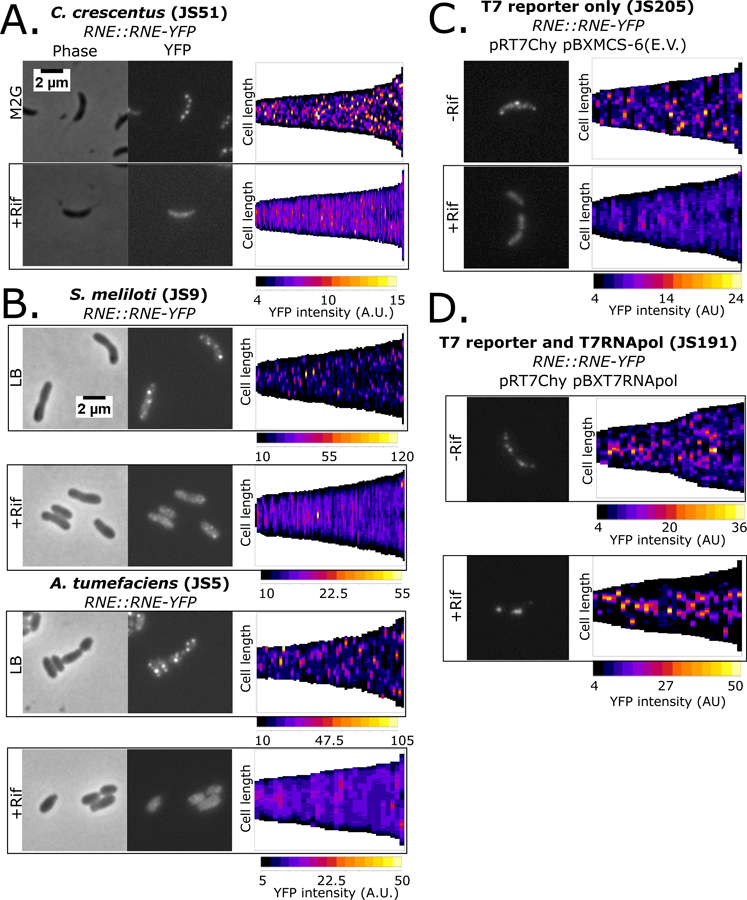
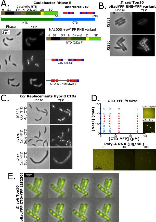
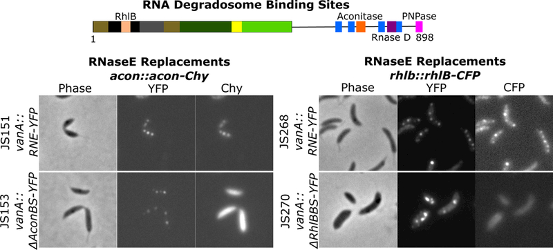
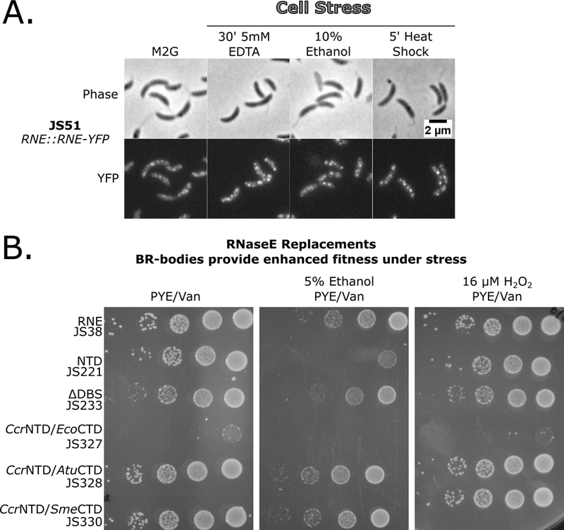
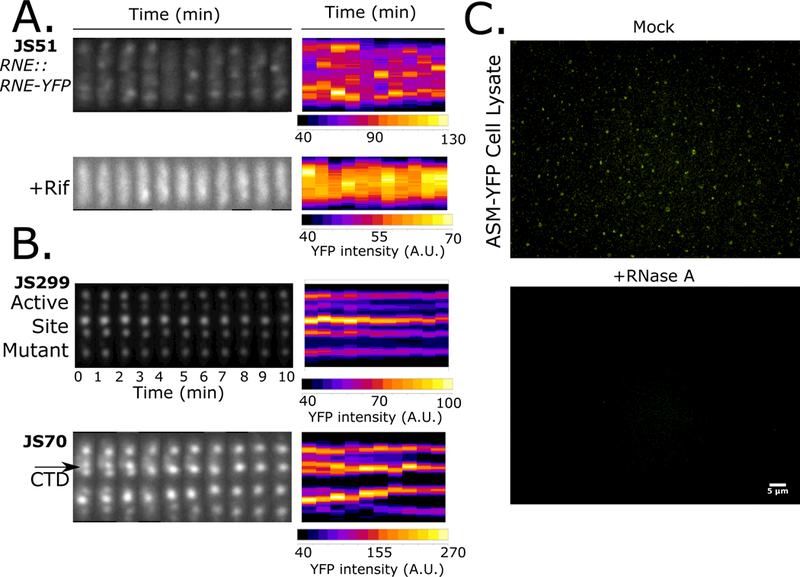
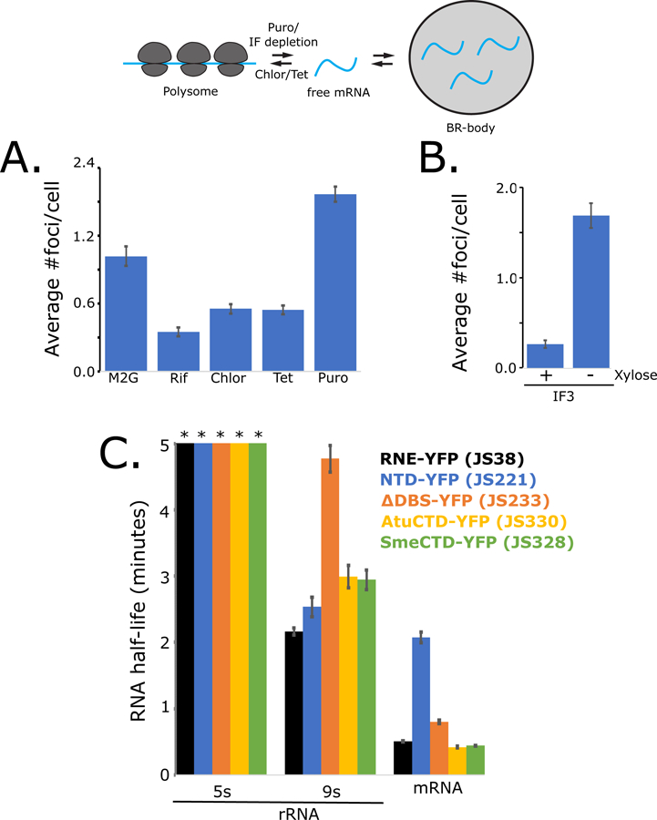
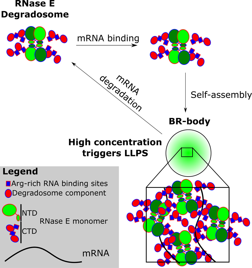
Ribonucleoprotein (RNP) granules play an important role in organizing eukaryotic mRNA metabolism via liquid-liquid phase separation (LLPS) of mRNA decay factors into membrane-less organelles in the cytoplasm. Here we show that the bacterium Caulobacter crescentus Ribonuclease (RNase) E assembles RNP LLPS condensates that we term bacterial RNP-bodies (BR-bodies), similar to eukaryotic P-bodies and stress granules. RNase E requires RNA to assemble a BR-body, and disassembly requires RNA cleavage, suggesting BR-bodies provide localized sites of RNA degradation. The unstructured C-terminal domain of RNase E is both necessary and sufficient to assemble the core of the BR-body, is functionally conserved in related α-proteobacteria, and influences mRNA degradation. BR-bodies are rapidly induced under cellular stresses and provide enhanced cell growth under stress. To our knowledge, Caulobacter RNase E is the first bacterial protein identified that forms LLPS condensates, providing an effective strategy for subcellular organization in cells lacking membrane-bound compartments.

Keywords: P-bodies; RNA degradation; RNase E; bacteria; liquid-liquid phase separation; mRNA decay; stress granules.


Conflict of interest statement

Declaration of Interests

The authors declare no competing interests exist.

Figures

Figure 1.
Figure 1.
α-proteobacterial RNase E assembles mRNA dependent foci. A.) Localization patterns for RNase E – YFP strain grown in M2G minimal media. Demograph displays the YFP intensity as a heat map across the long axis of single cells as vertical slices arranged by cell length (generated with microbeJ). B.) Localization patterns for log phase Sme 1021 or Atu C58 RNase E – YFP strains. C.) Localization patterns for Ccr cells expressing RNase E – YFP containing a T7 polymerase driven mCherry reporter plasmid (pRT7Chy) and a pBX empty vector plasmid (pBXMCS-6). D.) Localization patterns for Ccr cells expressing RNase E – YFP containing a pBX plasmid with rifR T7 polymerase (pBXT7RNApol) and a T7 driven mCherry reporter plasmid (pRT7Chy).
Figure 2.
Figure 2.
α-proteobacterial RNase E CTD is required for BR-body assembly into liquid-liquid phase separated condensates. A.) Domain architecture for the Ccr RNase E protein (Hardwick et al., 2011). The catalytic N-terminal domain (NTD, 1–450 amino acids) and disordered C-terminal domain (CTD, 451–898) are highlighted. H= RNase H domain, S1= ribosomal protein S1 domain, 5′P = 5′ monophosphate sensor domain, DNaseI= Catalytic region, Zn = Zinc link, SD= small domain, and AR= Arginine rich RNA binding sites. Positive and negative charged patches are indicated with red and blue boxes. Subcellular localization patterns in Ccr of the YFP tagged mutants of RNase E as indicated. B.) Heterologous subcellular localization patterns of Ccr RNase E in Eco cells with the YFP tagged mutants of RNase E as indicated. C.) RNase E CTD domain fusions with the CTD domains from Atu, Sme, or Eco fused to the Ccr NTD in RNase E replacement strains. D.) Phase diagram of purified Ccr RNase E CTD-YFP incubated at the indicated concentrations of protein and NaCl. LLPS condensate phase boundary is indicated by red circles. Right, condensates were formed with addition of the indicated concentrations of poly-A RNA at 100mM NaCl and 12.4 µM CTD-YFP. E.) Time-lapse images of JS230 showing condensate droplet fusion. Images were taken at 1′ intervals and the white arrows indicate the two BR-bodies that fuse.
Figure 3.
Figure 3.
RNase E recruits degradosome components into BR-bodies. Ccr RNase E protein with the RNA degradosome binding sites highlighted (Hardwick et al., 2011; Voss et al., 2014). Replacement strains harboring YFP tagged (RNE= full length, ΔAconBS=aconitase binding site deletion, ΔRhlBBS=RhlB binding site deletion) variants under control of the vanillate promoter and a natively tagged copy of acon-Chy or RhlB-CFP.
Figure 4.
Figure 4.
BR-body assembly is increased under specific stresses and provides increased stress tolerance. A.) Cells containing the RNase E-YFP fusion were subjected to cell stresses as indicated, gently mixed, spotted on the agarose pad, and immediately imaged. B.) BR-bodies provide enhanced survival under cell stress independent of degradosome formation. RNase E Replacement strains were grown under replacement conditions, diluted to the same OD600, and 1:10 serial dilutions were plated on PYE plates containing the selectable markers and indicated stress.
Figure 5.
Figure 5.
BR-bodies are dynamic and require RNA cleavage for disassembly. A.) Ccr RNase E-YFP localization patterns were monitored by time-lapse microscopy. All images were taken in 1′ intervals. A representative time-lapse from a cell is shown for each condition with the straightened cell images placed side by side (kymograph heat map generated via microbeJ). B.) Time lapse was performed in the catalytically inactive Ccr ASM and CTD replacement strains. Black arrow indicates a BR-body fusion event. C.) Lysates of the ASM-YFP replacement strain were prepared in a replacement strain. Lysates were then either treated with 0.1mg/mL RNase A or mock treated and imaged.
Figure 6.
Figure 6.
BR-bodies compete with ribosomes for untranslated mRNAs and affect mRNA turnover. A.) Number of RNase E-YFP foci per cell measured upon different translation inhibitor treatments. Error bars represent standard error. B.) Number of RNase E-YFP foci per cell measured upon depletion of translation initiation factor. Error bars represent standard error. C.) qRT-PCR measure of RNA half-life. RNA samples were taken after rifampicin treatment and measured by qRT-PCR using primers for the mature 5s rRNA, 9s pre-rRNA, and RNase E 5′ UTR of the Ccr RNase E replacement strains indicated. Error bars represent standard error of at least three replicates.
Figure 7.
Figure 7.
Model of Ccr RNase E BR-body assembly and disassembly. Free RNase E tetramers scaffold the RNA degradosome protein complex. In the presence of untranslated mRNA, RNA degradosomes can bind to the mRNA, increasing local concentration and causing liquid-liquid phase condensation from self-assembly of the disordered alternating charged-blocks. High local concentrations of RNase E accelerates mRNA degradation, facilitating dissolution from the BR-body.

References

    1. Aguirre AA, Vicente AM, Hardwick SW, Alvelos DM, Mazzon RR, Luisi BF, and Marques MV (2017). Association of the Cold Shock DEAD-Box RNA Helicase RhlE to the RNA Degradosome in Caulobacter crescentus. J. Bacteriol 199. - PMC - PubMed
    1. Ait-Bara S, and Carpousis AJ (2015). RNA degradosomes in bacteria and chloroplasts: classification, distribution and evolution of RNase E homologs. Mol. Microbiol 97, 1021–1135. - PubMed
    1. Banani SF, Lee HO, Hyman AA, and Rosen MK (2017). Biomolecular condensates: organizers of cellular biochemistry. Nat. Rev. Mol. Cell Biol 18, 285–298. - PMC - PubMed
    1. Barry RM, Bitbol AF, Lorestani A, Charles EJ, Habrian CH, Hansen JM, Li HJ, Baldwin EP, Wingreen NS, Kollman JM, et al. (2014). Large-scale filament formation inhibits the activity of CTP synthetase. eLife 3, e03638. - PMC - PubMed
    1. Bayas CA, Wang J, Lee MK, Schrader JM, Shapiro L, and Moerner WE (2018). Spatial organization and dynamics of RNase E and ribosomes in Caulobacter crescentus Proceedings of the National Academy of Sciences, In Press. - PMC - PubMed

Publication types

MeSH terms

LinkOut - more resources