. 2018 Aug 24:11:296.
doi: 10.3389/fnmol.2018.00296. eCollection 2018.

Anxiety Specific Response and Contribution of Active Hippocampal Neural Stem Cells to Chronic Pain Through Wnt/β-Catenin Signaling in Mice

Affiliations

Anxiety Specific Response and Contribution of Active Hippocampal Neural Stem Cells to Chronic Pain Through Wnt/β-Catenin Signaling in Mice

Youyi Zhao et al. Front Mol Neurosci. .

Abstract

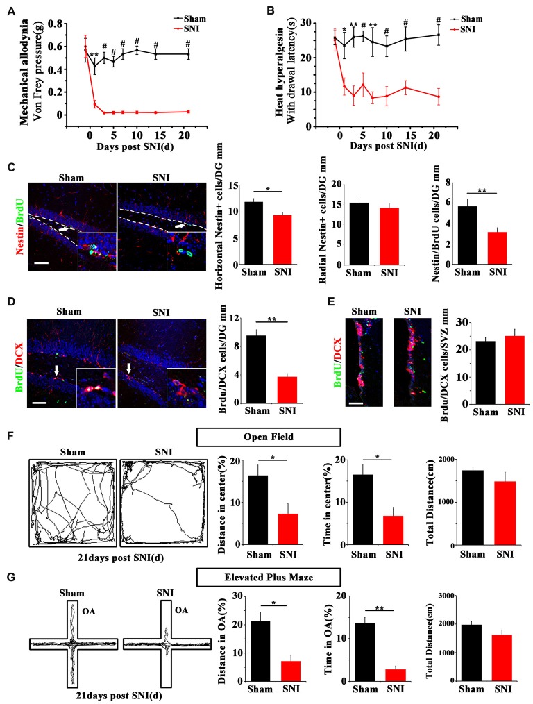
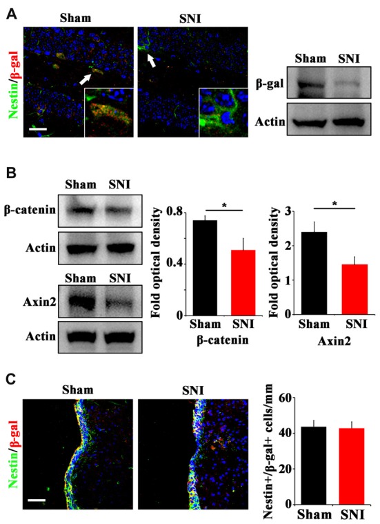
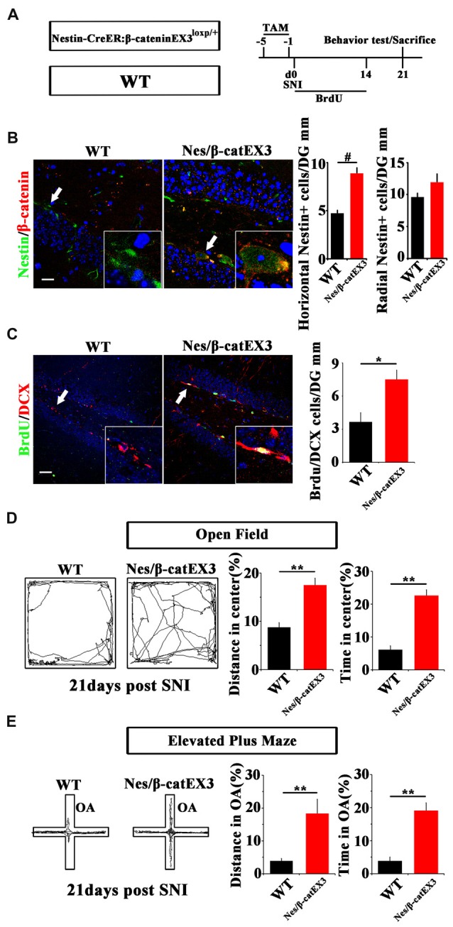
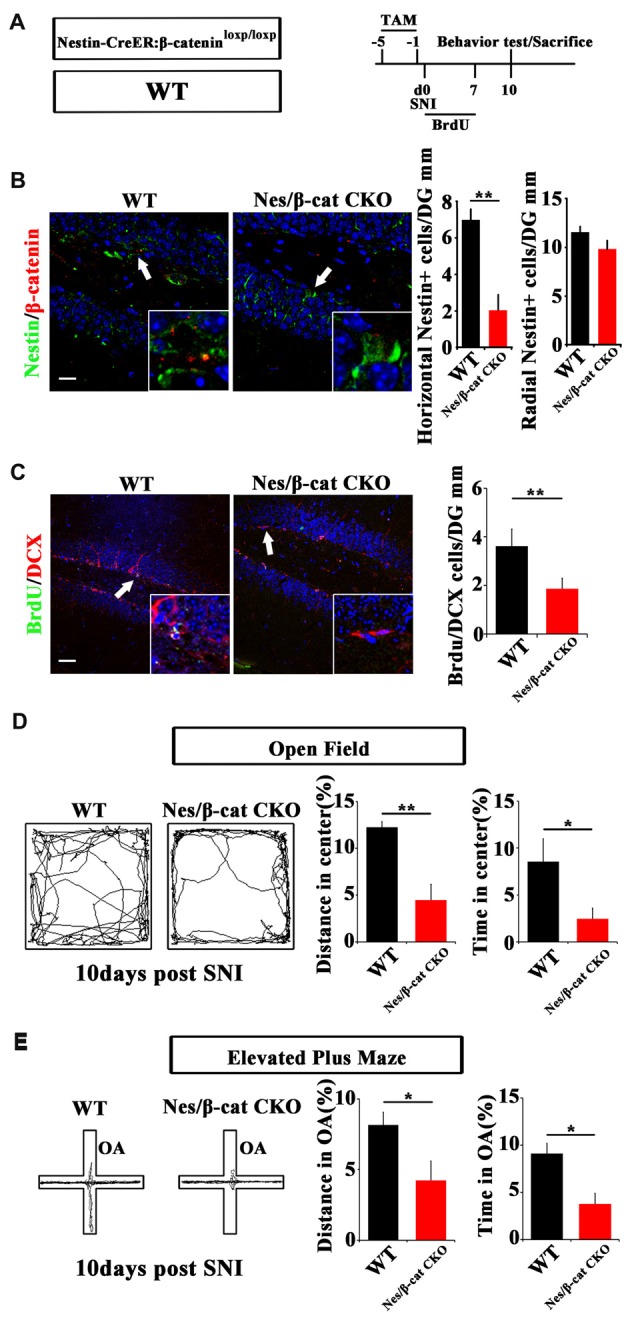
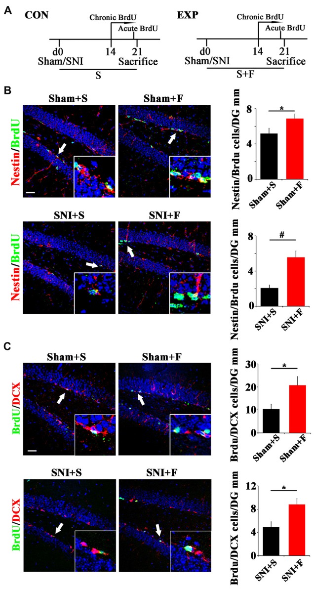
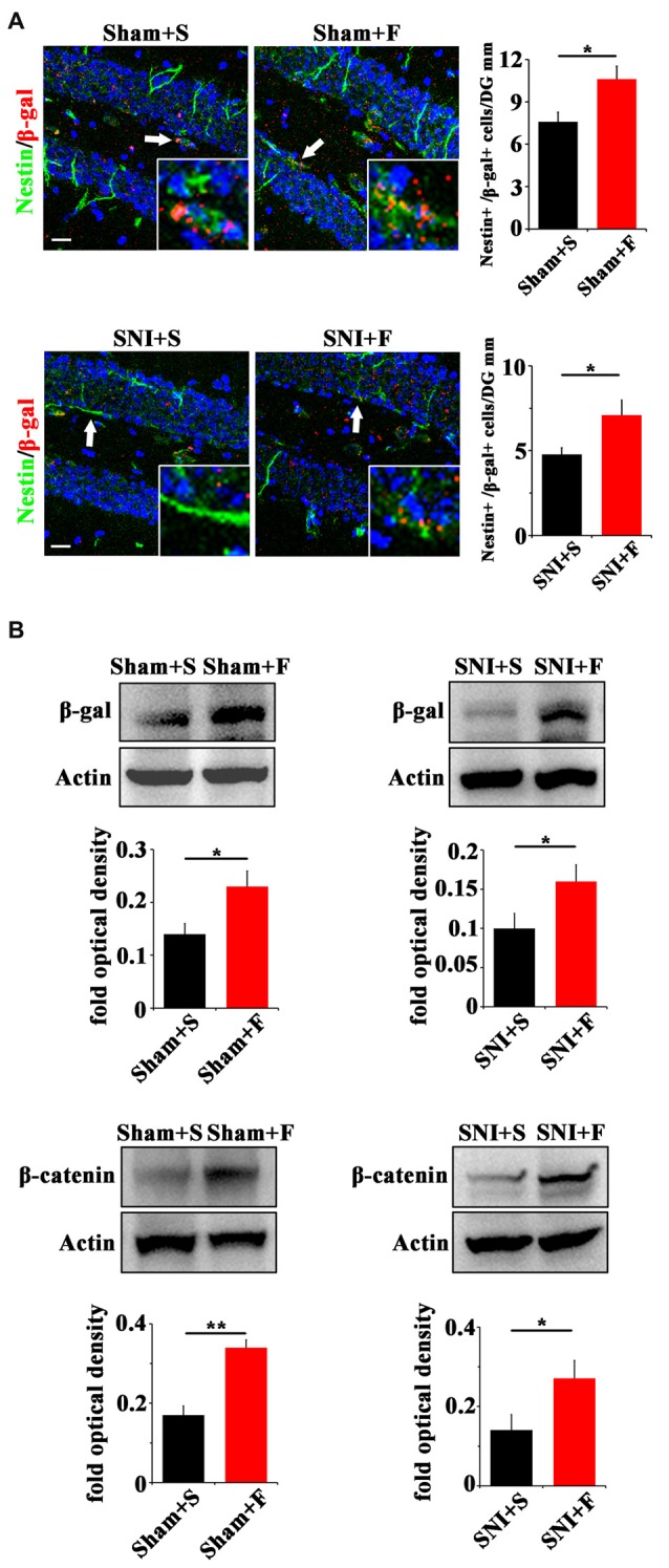
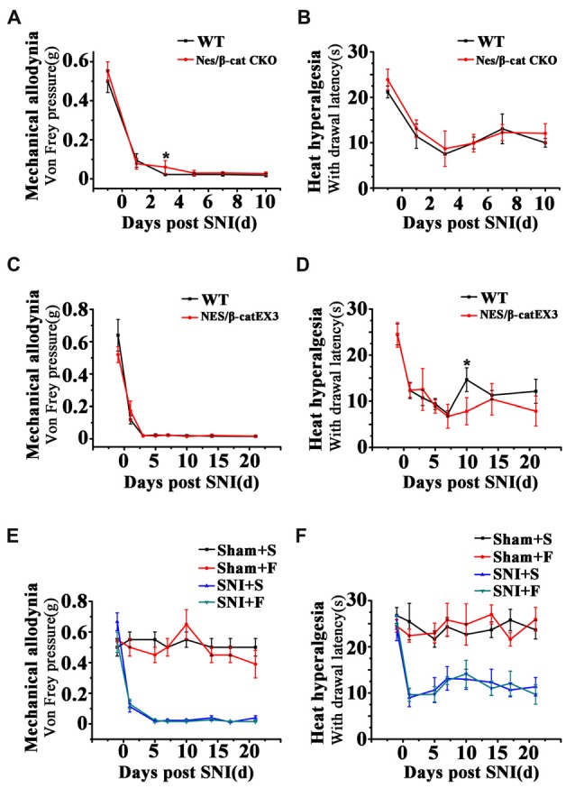
Chronic pain usually results in persistent anxiety, which worsens the life quality of patients and complicates the treatment of pain. Hippocampus is one of the few brain regions in many mammalians species which harbors adult neural stem cells (NSCs), and plays a key role in the development and maintenance of chronic anxiety. Recent studies have suggested a potential involvement of hippocampal neurogenesis in modulating chronic pain. Whether and how hippocampal NSCs are involved in the pain-associated anxiety remains unclear. Here, we report that mice suffering persistent neuropathic pain showed a quick reduction of active NSCs in the ventral dentate gyrus (vDG), which was followed by the decrease of neurogenesis and appearance of anxiety. Wnt/β-catenin signaling, a key pathway in sustaining the active status of NSCs was suppressed in the vDG of mice suffering chronic pain. Depleting β-catenin by inducible Nestin-Cre significantly reduced the number of active NSCs and facilitated anxiety development, while expressing stabilized β-catenin amplified active NSCs and alleviated anxiety, indicating that Wnt activated NSCs is required for anxiety development under chronic pain. Treatment with Fluoxetine, the most widely used anxiolytic in clinic, significantly increased the proliferation of active NSCs and enhanced Wnt signaling. Interestingly, both β-catenin manipulation and Fluoxetine treatment had no significant effects on the pain thresholds. Therefore, our data demonstrated an anxiety-specific response and contribution of activated NSCs to chronic pain through Wnt/β-catenin signaling, which may be targeted for treating chronic pain- or other diseases-associated anxiety.

Keywords: Wnt/β-catenin signaling; adult neural stem cell; anxiety; chronic pain; hippocampus.


Figures

Figure 1
Figure 1
Effects of chronic pain on the activation of neural stem cells (NSCs) in ventral hippocampus and the appearance of anxiety. (A) Pain threshold for mechanical allodynia after spared nerve injury (SNI). (B) Pain threshold for thermal hyperalgesia after SNI. Notice the persistence of chronic pain to at least 21 days after SNI. (C) Double-immunostaining of Nestin/BrdU and the quantification at 7 dpi after acute BrdU incorporation. Notice the decrease of horizontal Nestin-positive cells, which were the major BrdU-labeled cells in SNI-treated mice. (D) Double-immunostaining and quantification of DCX/BrdU-positive cells in ventral dentate gyrus (vDG) at 21 dpi. (E) Double-immunostaining and quantification of DCX/BrdU-positive cells in subventricular zone (SVZ) at 21 dpi. Notice the decrease of DCX/BrdU-positive cells in vDG and no change of DCX/BrdU-positive cells in SVZ between sham and SNI-treated mice. (F) Open field assay of sham-injured and SNI mice. (G) Elevated plus maze test of sham-injured and SNI mice. Notice the decrease of movements of SNI-treated mice in the open field and open arm. Bars = 50 μm in (C–E). OA, open arm. Inserts in (C,D) are magnified typical double-stained cells, which were pointed by arrows. Dashed lines in (C) showed the basal line along which quantification made. Values represent mean ± SE. One-way analysis of variance (ANOVA) analysis with Dunnett’s post hoc test was performed in (A,B). Unpaired, two tailed Student’s t-tests were performed in (C–G). *P < 0.05, **P < 0.01, #P < 0.001.
Figure 2
Figure 2
Effects of chronic pain on Wnt activity in ventral hippocampus. (A) Double-immunostaining of Nestin/β-gal and Western-blotting of β-gal in the ventral hippocampus of sham-injured and SNI-treated Topgal mice at 21 dpi. (B) Western-blotting of β-catenin and Axin2 in the ventral hippocampus of sham-injured and SNI-treated mice at 21 dpi. (C) Double-immunostaining and quantification of Nestin/β-gal in the SVZ of sham-injured and SNI-treated Topgal mice at 21 dpi. Inserts in (A) are magnified typical Nestin/β-gal-positive cells in each group, which were pointed by arrows. Bars = 25 μm in (A) and 50 μm in (C). Values represent mean ± SE. Unpaired, two tailed Student’s t-tests were performed in (B,C). *P < 0.05.
Figure 3
Figure 3
Effects of expressing stabilized β-catenin in adult neural progenitors on hippocampal neurogenesis and pain-associated anxiety. (A) Schematic drawing of the experimental design. (B) Double-immunostaining of Nestin/β-catenin and quantification of Nestin-positive cells SNI-treated wild-type (WT) or Nestin-β-catEX3 mice. Notice the enhanced expression of β-catenin and the increase of horizontal Nestin-positive cells in the hippocampus of Nestin-β-catEX3 mice. (C) Double-immunostaining and quantification of BrdU/DCX in SNI-treated WT or Nestin-β-catEX3 mice. (D,E) Open field and elevated plus maze tests of SNI-treated WT or Nestin-β-catEX3 mice at 21 dpi. Notice the alleviation of anxiety in Nestin-β-catEX3 mice with chronic pain. OA, open arm. Inserts in (B,C) are magnified typical double-stained cells in each group, which were pointed by arrows. Bars = 25 μm in (B) and 50 μm in (C). Values represent mean ± SE. Unpaired, two tailed Student’s t-tests were performed in (B,C). *P < 0.05, **P < 0.01, #P < 0.001.
Figure 4
Figure 4
Effects of ablating β-catenin in adult neural progenitors on hippocampal neurogenesis and pain-associated anxiety. (A) Schematic drawing of the experimental design. (B) Double-immunostaining of Nestin/β-catenin and quantification of Nestin-positive cells in SNI-treated WT or Nestin-β-cat CKO mice at 10 dpi. Notice the decrease of β-catenin immunoreactivity and radial Nestin-positive cells in the hippocampus of Nestin-β-cat CKO mice. (C) Double-immunostaining and quantification of BrdU/DCX in SNI-treated WT or Nestin-β-cat CKO mice at 10 dpi. (D,E) Open field and elevated plus maze tests of SNI-treated WT or Nestin-β-cat CKO mice at 10 dpi. Notice the appearance of anxiety in Nestin-β-cat CKO mice at 10 dpi. OA, open arm. Inserts in (B,C) are magnified typical double-stained cells in each group, which were pointed by arrows. Bars = 25 μm in (B) and 50 μm in (C). Values represent mean ± SE. Unpaired, two tailed Student’s t-tests were performed in (B–E). *P < 0.05, **P < 0.01.
Figure 5
Figure 5
Effects of Fluoxetine treatment on the NSC proliferation and neurogenesis in hippocampus. (A) Schematic drawing of the experimental design. (B) Double-immunostaining of BrdU/Nestin in sham mice treated with saline (Sham+S), sham mice treated with Fluoxetine (Sham+F), SNI mice treated with saline (SNI+S), and SNI mice treated with Fluoxetine (SNI+F). Notice that Fluoxetine treatment significantly increased the number of BrdU/Nestin-positive cells in both sham and SNI-treated mice after acute BrdU incorporation, as compared to corresponding saline controls. (C) Double-immunostaining of BrdU/DCX in sham mice treated with saline (Sham+S), sham mice treated with Fluoxetine (Sham+F), SNI mice treated with saline (SNI+S), and SNI mice treated with Fluoxetine (SNI+F). Notice that Fluoxetine treatment significantly increased the number of BrdU/DCX-positive cells after 7 days’ successive BrdU administration. CON, control group. EXP, experimental group. S, saline. F, Fluoxetine. Inserts in (B,C) are typical double-stained cells in each group, which were pointed by arrows. Bars = 50 μm. Values represent mean ± SE. Unpaired, two tailed Student’s t-tests were performed in (B,C). *P < 0.05, #P < 0.001.
Figure 6
Figure 6
Effects of Fluoxetine treatment on the Wnt activity in hippocampus. (A) Double-immunostaining of Nestin/β-gal in sham mice treated with saline (Sham+S), sham mice treated with Fluoxetine (Sham+F), SNI mice treated with saline (SNI+S), and SNI mice treated with Fluoxetine (SNI+F) at 21 dpi. Notice the significant increase of radial Nestin-positive cells in Fluoxetine treated mice. (B) Western-blotting of β-gal and β-catenin in Topgal mice with the following treatments: sham injury plus saline (Sham+S), sham injury plus Fluoxetine (Sham+F), SNI plus saline (SNI+S) and SNI plus Fluoxetine (SNI+F). Notice that Fluoxetine treatment significantly increased the expression level of β-gal and β-catenin in both sham and SNI-treated mice, as compared to corresponding saline controls. S, saline. F, Fluoxetine. Inserts in (A) are typical double-stained cells in each group, which were pointed by arrows. Bars = 50 μm. Values represent mean ± SE. Unpaired, two tailed Student’s t-tests were performed in (A,B). *P < 0.05, **P < 0.01.
Figure 7
Figure 7
Effects of Wnt modulation and Fluoxetine treatment on pain thresholds. (A,B) Effects of ablation of β-catenin in hippocampal neural progenitors on mechanical and thermal withdraw thresholds of SNI treated mice. (C,D) Effects of expressing stabilized β-catenin in hippocampal neural progenitors on mechanic and thermal withdraw thresholds of SNI treated mice. (E,F) Effects of Fluoxetine treatment on mechanic and thermal withdraw thresholds of sham injured and SNI treated mice. S, saline. F, Fluoxetine. Notice the no change of pain thresholds in mice with β-catenin manipulation or Fluoxetine treatment. Values represent mean ± SE. One-way ANOVA analysis with Dunnett’s post hoc test was performed in (A–F). *P < 0.05.

Similar articles

Cited by

References

    1. Antonelli F., Casciati A., Tanori M., Tanno B., Linares-Vidal M. V., Serra N., et al. . (2018). Alterations in morphology and adult neurogenesis in the dentate gyrus of patched1 heterozygous mice. Front. Mol. Neurosci. 11:168. 10.3389/fnmol.2018.00168 - DOI - PMC - PubMed
    1. Apkarian A. V., Mutso A. A., Centeno M. V., Kan L., Wu M., Levinstein M., et al. . (2016). Role of adult hippocampal neurogenesis in persistent pain. Pain 157, 418–428. 10.1097/j.pain.0000000000000332 - DOI - PMC - PubMed
    1. Benedetti M., Merino R., Kusuda R., Ravanelli M. I., Cadetti F., Dos Santos P., et al. . (2012). Plasma corticosterone levels in mouse models of pain. Eur. J. Pain 16, 803–815. 10.1002/j.1532-2149.2011.00066.x - DOI - PubMed
    1. Bliss T. V., Collingridge G. L., Kaang B. K., Zhuo M. (2016). Synaptic plasticity in the anterior cingulate cortex in acute and chronic pain. Nat. Rev. Neurosci. 17, 485–496. 10.1038/nrn.2016.68 - DOI - PubMed
    1. Brault V., Moore R., Kutsch S., Ishibashi M., Rowitch D. H., McMahon A. P., et al. . (2001). Inactivation of the β-catenin gene by Wnt1-Cre-mediated deletion results in dramatic brain malformation and failure of craniofacial development. Development 128, 1253–1264. - PubMed

LinkOut - more resources