Review
. 2018 Aug 24:9:1147.
doi: 10.3389/fphys.2018.01147. eCollection 2018.

Sense and Insensibility - An Appraisal of the Effects of Clinical Anesthetics on Gastropod and Cephalopod Molluscs as a Step to Improved Welfare of Cephalopods

Affiliations
Review

Sense and Insensibility - An Appraisal of the Effects of Clinical Anesthetics on Gastropod and Cephalopod Molluscs as a Step to Improved Welfare of Cephalopods

William Winlow et al. Front Physiol. .

Abstract

Recent progress in animal welfare legislation stresses the need to treat cephalopod molluscs, such as Octopus vulgaris, humanely, to have regard for their wellbeing and to reduce their pain and suffering resulting from experimental procedures. Thus, appropriate measures for their sedation and analgesia are being introduced. Clinical anesthetics are renowned for their ability to produce unconsciousness in vertebrate species, but their exact mechanisms of action still elude investigators. In vertebrates it can prove difficult to specify the differences of response of particular neuron types given the multiplicity of neurons in the CNS. However, gastropod molluscs such as Aplysia, Lymnaea, or Helix, with their large uniquely identifiable nerve cells, make studies on the cellular, subcellular, network and behavioral actions of anesthetics much more feasible, particularly as identified cells may also be studied in culture, isolated from the rest of the nervous system. To date, the sorts of study outlined above have never been performed on cephalopods in the same way as on gastropods. However, criteria previously applied to gastropods and vertebrates have proved successful in developing a method for humanely anesthetizing Octopus with clinical doses of isoflurane, i.e., changes in respiratory rate, color pattern and withdrawal responses. However, in the long term, further refinements will be needed, including recordings from the CNS of intact animals in the presence of a variety of different anesthetic agents and their adjuvants. Clues as to their likely responsiveness to other appropriate anesthetic agents and muscle relaxants can be gained from background studies on gastropods such as Lymnaea, given their evolutionary history.

Keywords: behavior; cephalopods; clinical anesthetics; gastropods; identified neurons.


Figures

FIGURE 1
FIGURE 1
Dorsal view of the central nervous system of Lymnaea stagnalis (L.) with the exception of the paired buccal ganglia. The diagram shows locations of individual neurons (black), cell groups and clusters (clear) identified in the text. LCGC and RCGC, left and right cerebral giant cells; RPeD1, right pedal dorsal cell 1; LPeD1, left pedal dorsal cell 1 VD1 and VD4, visceral dorsal cells 1 and 4; RPD1 and RPD2, right parietal; dorsal cells 1 and 2; Ip3I, input 3 interneuron; A gp, right parietal A group; M gp, visceral M group; LPeA and RPeA, left and right pedal A group cells; PeI, pedal I group cells. A, anterior; P, posterior; L, left; R. right. L.C.G. and R.C.G, left and right cerebral ganglia; L.Pe.G. and R.Pe.G., left and right pedal ganglia; L.Pl.G. and R.Pl.G., left and right pleural ganglia; L.P.G. and R.P.G., left and right parietal ganglia: V.G., median visceral ganglion; Idb., lateral dorsal body; II., lateral lobe; St, statocyst. (1) cerebro-buccal connective: (2) superior labial nerve; (3) median labial nerve; (4) penis nerve; (5) tentacle nerve; (6) optic nerve; (7) nuchal nerve; (8) left parietal nerve; (9) cutaneous pallial nerve; (10) intestinal nerve; (11) anal nerve; (12) genital nerve; (13) right internal parietal nerve; (14) right external parietal nerve; (15) inferior cervical nerve; (16) superior cervical nerve; (17) columellar nerve; (18) superior pedal nerve; (19) inferior pedal nerve; (20) medial pedal nerve; (21) dorsal pedal commissure; (22) ventral pedal commissure; (23) medial columellar nerve; (24) cerebro-pedal connective: (25) pedal-pleural connective.
FIGURE 2
FIGURE 2
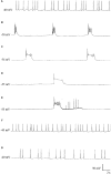
The effect of Pentobarbital on the spontaneous firing pattern and frequency of an A group neuron. (A) control; (B) 1 mM pentobarbital after 6 min; (C) 1 mM pentobarbital after 30 min; (D) 2 mM pentobarbital after 6 min; (E) 10 min wash out prior to quiescence and (F) 30 min wash out and (G) 60 min wash out. Membrane potential increased in response to pentobarbital, and decreased as continuous wash out of pentobarbital proceeded. (From Moghadam, 1996 – Reproduced under the Creative Commons License).
FIGURE 3
FIGURE 3
Dose dependent depression of peak current and endpulse current in a PeI cluster cell of Lymnaea stagnalis. The Cell was held at a holding potential of –50 mV and 180 ms long depolarizing pulses were applied in 10 mV increments in control (snail saline), 0.5, 1.0, 2.0, and 4.0% halothane. There is a very clear reduction of calcium channel current which is concentration-dependent. The peaks of the I-V curves are not shifted by halothane. (From Yar and Winlow, 2016 – Reproduced under the Creative Commons License).
FIGURE 4
FIGURE 4
Suppression of potassium currents by both isoflurane (A) and sodium pentobarbital (B) in cultured pedal I cluster neurons of Lymnaea, using the whole cell voltage clamp technique. (From Moghadam, 1996 - Reproduced under the Creative Commons License).
FIGURE 5
FIGURE 5
Ratiofluorimetric images showing that Halothane raises intracellular calcium concentration in the cultured Lymnaea neurone RPD2 loaded with the fluorescent Ca2+ indicator Fura 2. (A) Control in normal saline (101.18 nM [Ca2+]I); (B) 1 min after addition of 2% halothane (174.12 nM [Ca2+]I); (C) 5 min after 2% halothane (287.06 nM [Ca2+]I); (D) 10 min after washout of 2% halothane with normal saline (183.53 nM [Ca2+]I); (E) 1 min after 4% halothane (232.94 nM [Ca2+]I); (F) 5 min after 4% halothane (414.12 nM [Ca2+]I); (G) 15 min after washout of 4% halothane with normal saline (211.76 nM [Ca2+]I); (H) Fluorescence scale bar (From Ahmed, 1995 – Reproduced under the Creative Commons License).
FIGURE 6
FIGURE 6
Halothane modifies intracellular calcium concentration in RPeD1, (A) when vaporized into normal saline, (B) when vaporized into zero calcium saline (From Ahmed, 1995 – Reproduced under the Creative Commons License).
FIGURE 7
FIGURE 7
Diagram of the neurons making up the respiratory CPG of Lymnaea stagnalis. RPeD1 initiates the RCPG rhythm, VD4 drives inspiration and pneumostome closure, while the input 3 interneuron (Ip3I) drives expiration and pneumostome opening. Ovals are inhibitory synapses and arrows are excitatory synapses. RPeD1 has a biphasic synaptic connection to Ip3I (For further detail, see Syed et al., 1990).
FIGURE 8
FIGURE 8
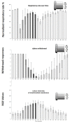
Illustration of behavioral criteria for anesthesia in Octopus vulgaris. Effects of isoflurane on respiratory rate (upper), siphon withdrawal (middle) and color intensity of the interbrachial membrane (lower) in Octopus vulgaris. Normalized respiratory rate as determined by the number of mantle contractions in 10 animals at 5 min intervals. The relative strength of siphon withdrawal in response to a touch stimulus (6 = strong, 4 = medium, 2 = low and 0 = none). The color intensity was measured using imageJ software and the RGB model, whereby a zero intensity (value 0) for each component gives the darkest color (no light = black) and full intensity for each component gives white (value 255). Thus the higher recorded value, the paler the interbrachial membrane and vice versa. After 50 min the seawater tank was refreshed and the isoflurane flow was switched off. Error bars are standard deviations. (Modified from Polese et al., 2014 – with permission from Journal of Aquatic Animal Health, 2014, Publisher John Wiley and Sons).

Similar articles

Cited by

References

    1. Ahmed I. A. (1995). Effects of General Anaesthetics and Other Pharmacological agents on Intracellular Calcium Levels in Identified Molluscan Neurons. Ph.D. thesis, University of Leeds, Leeds.
    1. Akaike N., Ito H., Nishi K., Oyama Y. (1982). Further analysis of inhibitory effects of propranolol and local anaesthetics on the calcium current in Helix neurones. Br. J. Pharmacol. 76 37–43. 10.1111/j.1476-5381.1982.tb09188.x - DOI - PMC - PubMed
    1. Alupay J. S., Hadjisolomou S. P., Crook R. J. (2014). Arm injury produces long-term behavioural and neural hypersensitivity in octopus. Neurosci. Lett. 558 137–142. 10.1016/j.neulet.2013.11.002 - DOI - PubMed
    1. Andrews P. L. R., Darmaillacq A. S., Dennison N., Gleadall I. G., Hawkins P., Messenger J. B., et al. (2013). The identification and management of pain, suffering and distress in cephalopods, including anaesthesia, analgesia and humane killing. J. Exp. Mar. Biol. Ecol. 447 46–64. 10.1016/j.jembe.2013.02.010 - DOI
    1. Andrews P. L. R., Tansey T. (1981). The effects of some anaesthetic agents in Octopus vulgaris. Comp. Biochem. Physiol. C Comp. Pharmacol. 70 241–247. 10.1016/0306-4492(81)90057-5 - DOI

LinkOut - more resources