. 2018 Aug 21;9(65):32478-32495.
doi: 10.18632/oncotarget.25980.

Zac1 regulates IL-11 expression in osteoarthritis

Affiliations

Zac1 regulates IL-11 expression in osteoarthritis

Chun-Lin Kuo et al. Oncotarget. .

Abstract

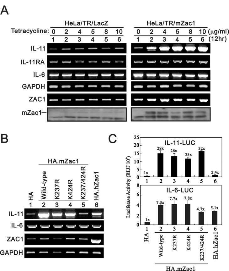
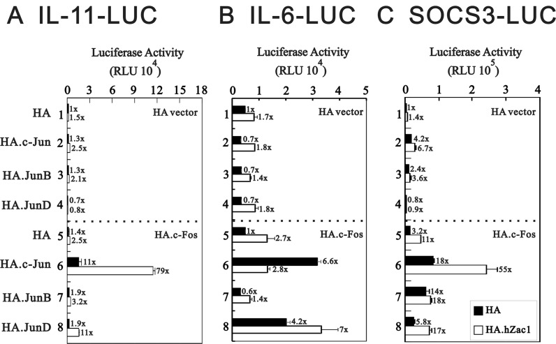
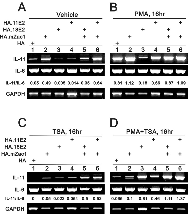
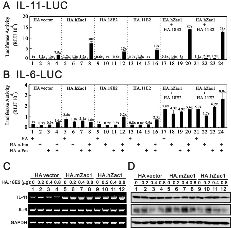
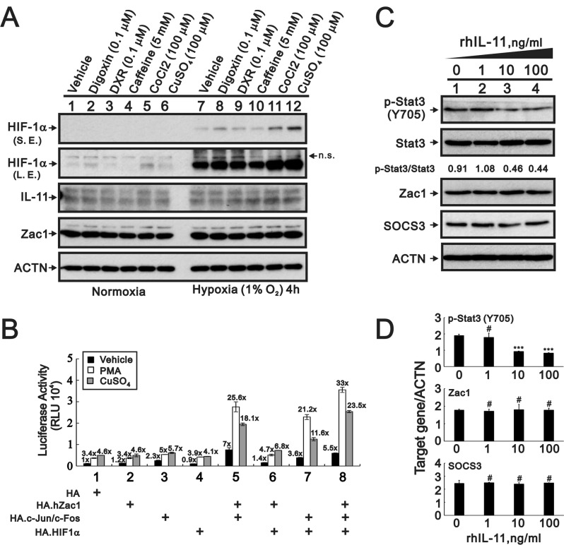
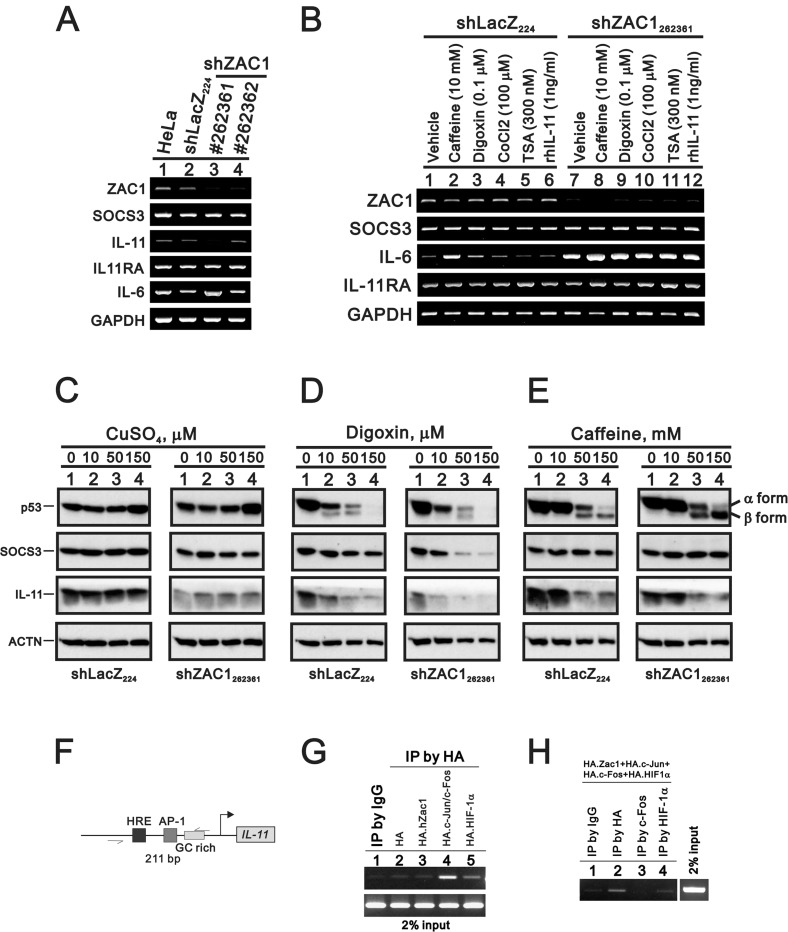
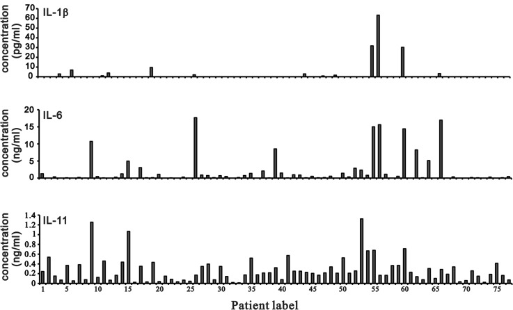
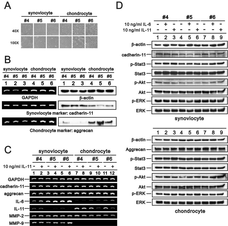
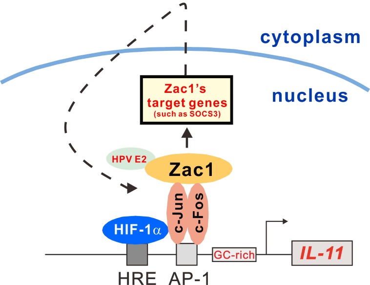
Interleukin (IL)-11, a member of the IL-6 family of cytokines, exerts pleiotropic effects under normal and various disease conditions. We assessed IL-11 expression regulation and the IL-11/IL-6 ratio in osteoarthritis (OA) to better guide clinical therapeutic decision-making. Our findings suggest that Zac1, a zinc finger protein that regulates apoptosis and cell cycle arrest, is a transcription factor regulating IL-11 expression. Zac1 overexpression or knockdown respectively induced or suppressed IL-11 expression in HeLa cells. Zac1 acted synergistically with AP-1, human papillomavirus E2, and hypoxia inducible factor 1 alpha (HIF1α). IL-11 expression under various conditions, including hypoxia or treatment with phorbol 12-myristate 13-acetate or copper sulfate. Recombinant IL-11-induced phosphorylation of signal transducer and activator of transcription 3 at tyrosine 705 was reduced in a dose-dependent manner in HeLa cells. Cross-talk between Zac1, IL-11, p53, and suppressor of cytokine signaling 3 was differentially affected by copper sulfate, digoxin, and caffeine. Finally, aggressive vs. conventional treatment of OA patients was primarily determined by IL-6 levels. However, we suggest that OA patients with higher IL-11 levels may respond well to conventional treatments, even in the presence of high IL-6.

Keywords: HeLa; IL-11; IL-6; osteoarthritis; zac1.


Conflict of interest statement

CONFLICTS OF INTEREST The authors declare that they have no conflicts of interest.

Figures

Figure 1
Figure 1. Effects of overexpressing Zac1 on the IL-11 mRNA and promoter in HeLa cells
(A) HeLa/TR/mZac1 or HeLa/TR-LacZ control cells were induced for 12 h using the indicated amounts of Dox. The cells were collected and subjected to RT-PCR analysis of IL-11, IL-6, IL-11Rα, Zac1, and GAPDH (loading control) mRNA expression, and to immunoblot analysis for detection of Zac1 protein. (B) HeLa cells were transiently transfected with 0.5 μg of the indicated pSG5.HA.Zac1 for 36 h. The cells were then collected and subjected to RT-PCR analysis of IL-11, IL-6, IL-11Rα, Zac1, and GAPDH (loading control) expression. (C) HeLa cells were transiently transfected with 0.5 μg of IL-11-LUC or IL-6-LUC along with 0.5 μg of the indicated pSG5.HA Zac1 for 36 h prior to use in luciferase reporter assays. The numbers above the columns indicate the luciferase activity relative to an index of 1 assigned to the reporter with empty vector. Results are representative of three independent experiments.
Figure 2
Figure 2. Effect of cross-talk between Zac1 and AP1 on the promoter activities of IL-11, IL-6 and SOCS3
HeLa cells were transiently transfected for 36 h with 0.5 μg of IL-11-LUC (A), IL-6-LUC (B), or SOCS3-LUC (C) along with 0.5 μg of pSG5.HA, pSG5.HA.c-Jun, pSG5.HA.JunB, and pSG5.HA.JunD in the absence and presence of 0.5 μg of pSG5.HA, pSG5.HA.c-Fos or pSG5.HA.hZac1. The transfectants were then harvested for luciferase reporter assays. The numbers above the columns (A–C) indicate the luciferase activity relative to an index of 1 assigned to the reporter with empty vector. Results are representative of three independent experiments.
Figure 3
Figure 3. The rescue effect of Zac1 on HPV E2 proteins suppresses IL-11 mRNA expression
HeLa cells were transiently transfected for 20 h with 0.5 μg of pSG5.HA, pSG5.HA.mZac1, pSG5.HA.11E2, and pSG5.HA.18E2 and then treated with vehicle (A), PMA (B), TSA (C), or PMA (D) for 16 h. The cells were then collected and subjected to RT-PCR analysis of IL-11, IL-6, and GAPDH (loading control) expression. The IL-11/IL-6 ratio is presented as fold change. Results are representative of three independent experiments.
Figure 4
Figure 4. HPV E2 protein within the Zac1/AP1 complex enhances IL-11 promoter activities
HeLa cells were transiently transfected for 36 h with 0.5 μg of IL-11-LUC (A) or IL-6-LUC (B) along with 0.5 μg of pSG5.HA, pSG5.HA.c-Jun, pSG5.HA.c-Fos, pSG5.HA.hZac1, pSG5.HA.11E2 or pSG5.HA.18E2. Cells were then harvested for luciferase reporter assays. The numbers above the columns (A and B) indicate the luciferase activity relative to an index of 1 assigned to the reporter with empty vector. HeLa cells were transiently transfected for 36 h with 0.5 μg of pSG5.HA, pSG5.HA.mZac1, pSG5.HA.hZac1, and pSG5.HA.18E2 along with 0.5 μg of pSG5.HA.18E2. The cells were then collected and subjected to RT-PCR analysis of IL11, IL-6, and GAPDH (loading control) mRNA expression (C) and immunoblot analysis of IL-11, IL-6, and ACTN (loading control) protein expression (D). Results are representative of three independent experiments.
Figure 5
Figure 5. Expression of IL-11 protein is modulated by cobalt chloride, copper sulfate, and hypoxia
(A) HeLa cells were cultured for 4 h in the presence of digoxin (0.1 µM), DXR (0.1 µM), caffeine (5 mM), CoCl2 (100 µM), or CuSO4 (100 µM). The cells were then collected and subjected to Western blot analysis for detection of HIF1α, IL-11, Zac1, and PCNA (loading control) expression. S.E.: shorter exposure, L.E.: longer exposure, n.s.: not specific band. (B) HeLa cells were transiently transfected with 0.5 μg of IL-11-LUC along with 0.5 μg of pSG5.HA, pSG5.HA.HIF-1α, pSG5.HA.c-Jun, or pSG5.HA.c-Fos in the presence of vehicle, PMA, or copper sulfate for 36 h. Cells were then harvested for luciferase reporter assays. The numbers above the columns indicate the luciferase activity relative to an index of 1 assigned to the reporter with empty vector and vehicle. (CD) HeLa cells were treated for 24 h with indicated amounts of rIL-11. (C) The cells were collected and subjected to immunoblot analysis for the detection of p-Stat3 (Y705), Stat3, Zac1, SOCS3, and ACTN (loading control) expression. (D) Quantitative analysis of proteins is presented as the mean ± S.D. of at least three independent experiments; #p > 0.05 and ***p < 0.001 vs the level of ACTN (Student’s t-test).
Figure 6
Figure 6. Effects of copper sulfate, digoxin, and caffeine on p53 and SOCS3 expression in ZAC1 knockdown cells
(A) Parental HeLa cells, an shLacZ control clone and two shZAC1 knockdown clones were collected and subjected to RT-PCR analysis of ZAC1, SOCS3 IL-11, IL-6, IL-11Rα, and GAPDH (loading control) mRNA expression. (B) HeLa shLacZ and shZAC1262361 cells were treated for 20 h with caffeine (10 mM), digoxin (0.1 µM), CoCl2 (100 µM), TSA (300 nM), or rIL-11 (1 ng/ml), after which they were subjected to RT-PCR analysis of ZAC1, SOCS3 IL-11, IL-6, IL-11Rα, and GAPDH (loading control) mRNA expression. HeLa shLacZ and shZAC1262361 cells were treated for 20 h with indicated amounts of copper sulfate (C), digoxin (D), or caffeine (E) and subjected to immunoblot analysis for the detection of p53, SOCS3, IL11, and ACTN (loading control) protein expression. (F) Schematic representation of the hypoxia responsive element (HRE), AP-1 binding element, and GC-rich region of the IL-11 promoter. (GH) HeLa cells were transiently transfected for 36 h with 4 μg of pSG5HA vector, pSG5HA.Zac1, pSG5HA.c-Jun+pSG5HA.c-Fos, or pSG5HA.HIF-1α (G) or with 2 μg of pSG5HA.Zac1, pSG5HA.c-Jun+pSG5HA.c-Fos, and pSG5HA.HIF-1α (H). Cell lysates were immunoprecipitated using anti-IgG and anti-HA antibodies (G) or with anti-IgG, anti-HA, anti-c-Fos, or anti-HIF-1α antibody (H) and then subjected to PCR analysis. Results are representative of three independent experiments.
Figure 7
Figure 7. IL-1β, IL-6, and IL-11 levels in synovial fluid from OA patients
IL-1β, IL-6, and IL-11 levels were measured in samples of synovial fluid from OA patients using a BD™ CBA Human Soluble Protein Flex Set System.
Figure 8
Figure 8. Characteristics of primary synoviocytes and chondrocytes
Synoviocytes and chondrocytes from OA patients were observed under a light microscope (A) and subjected to RT-PCR analysis of cadherin-11, aggrecan, and GAPDH (loading control) mRNA expression and immunoblot analysis of cadherin-11, aggrecan, and β-actin (loading control) protein expression (B). (C, D) Synoviocytes and chondrocytes were treated with 10 ng/ml rIL-11 for 20 h, after which the cells were subjected to RT-PCR analysis of cadherin-11, aggrecan, IL-6, IL-11, MMP-2, MMP-9, and GAPDH (loading control) mRNA expression (C) and immunoblot analysis of cadherin-11 (for synoviocytes), aggrecan (for chondrocytes), p-Stat3, Stat3, p-ERK, ERK, p-Akt, Akt, and β-actin (loading control) protein expression (D). Results are representative of three independent experiments.
Figure 9
Figure 9. Schematic representation of the HRE and AP-1 binding element and the GC-rich region of the IL-11 promoter
Transcription factor Zac1 may not only induce transcription of target genes, such as SOCS3, it may also bind to host cellular proteins involved into the IL-11 gene regulation, such as HPV E2. Zac1 might also act as a transcriptional factor coactivator for AP-1 and HIF-1α to form induction complexes on the IL-11 promoter in HeLa cells.

Similar articles

Cited by

References

    1. Lokau J, Agthe M, Flynn CM, Garbers C. Proteolytic control of Interleukin-11 and Interleukin-6 biology. Biochim Biophys Acta. 2017;1864:2105–17. - PubMed
    1. Schaper F, Rose-John S. Interleukin-6: Biology, signaling and strategies of blockade. Cytokine Growth Factor Rev. 2015;26:475–87. - PubMed
    1. Garbers C, Scheller J. Interleukin-6 and interleukin-11: same same but different. Biol Chem. 2013;394:1145–61. - PubMed
    1. Naka T, Nishimoto N, Kishimoto T. The paradigm of IL-6: from basic science to medicine. Arthritis Res. 2002;4:S233–42. - PMC - PubMed
    1. Muraki S, Oka H, Akune T, Mabuchi A, En-yo Y, Yoshida M, Saika A, Suzuki T, Yoshida H, Ishibashi H, Yamamoto S, Nakamura K, Kawaguchi H, et al. Prevalence of radiographic knee osteoarthritis and its association with knee pain in the elderly of Japanese population-based cohorts: the ROAD study. Osteoarthritis Cartilage. 2009;17:1137–43. - PubMed