

Pacemaker lead endocarditis with hiccups (Kalayci)
- PMID: 30197777
- PMCID: PMC6121350
- DOI: 10.22088/cjim.9.3.299
Pacemaker lead endocarditis with hiccups (Kalayci)
Abstract
Background: Lead-related infections that might develop after pacemaker implantation associated with high mortality and morbidity rates are challenging to manage and pose high-cost. Patients with lead-related infections usually present with fever, chills and fatigue and the treatment can be challenging unless the implant system is extracted.
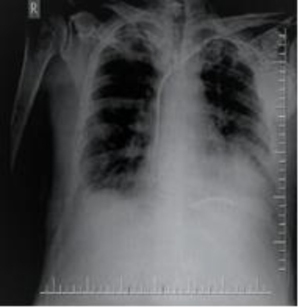
Case presentation: A 66-year old male patient who underwent dual chamber pacemaker and implantable cardioverter defibrillator was admitted to the emergency service with a six-week history of complaints of hiccups and fever. After a detailed investigation, lead-related infective endocarditis was the diagnosis. The patient was initiated on antibiotic therapy and lead extraction was performed.
Conclusions: Patients with signs of infection who underwent pacemaker implantation may present with atypical symptoms such as hiccup. In these cases, imaging, particularly echocardiography, should be performed as soon as possible and the localization of the pacemaker leads and signs of infective endocarditis should be investigated.
Keywords: Echocardiography; Hiccup; Infective endocarditis; Pacemaker lead.
Figures


References
-
- Kurtz SM, Ochoa JA, Lau E, et al. Implantation trends and patient profiles for pacemakers and implantable cardioverter defibrillators in the United States: 1993-2006. Pacing Clin Electrophysiol. 2010;33:705–11. - PubMed
-
- Voigt A, Shalaby A, Saba S. Continued rise in rates of cardiovascular implantable device infection in the United States: temporal trends and causative insights. Pacing Clin Electrophysiol. 2010;33:414–19. - PubMed
-
- Greenspon AJ, Patel JD, Lau E, et al. 16-year trends in the infection burden for pacemakers and implantable cardioverter-defibrillators in the United States 1993 to 2008. J Am Coll Cardiol. 2011;58:1001–6. - PubMed
-
- Yew KL. Infective endocarditis and the pacemaker: cardiac implantable electronic device infection. Med J Malaysia. 2012;67:618–9. - PubMed
Publication types
LinkOut - more resources
Full Text Sources